Friday, November 27, 2009

Various Causes of Acute Renal Failure

The cause and/or precipitating factor of acute renal failure (ARF) is always responsible for the effectiveness of therapy and supportive care techniques including hemodialysis. A rapid loss of renal function is exhibited through elevated levels of serum creatinine and blood urea due to fall in the clearance of these nitrogenous wastes by the kidneys in all cases of ARF. It has been observed that a loss of 50% of glomerular filtration rate (GFR) leads to significant elevation of the level of creatinine in the blood with a decrease in the urine output (oliguria). There could be three types of causes and implicating factors of acute renal failure: 1) Pre-renal, 2) Renal and 3) Post-renal. In pre-renal type ARF causes are the physiological factors or conditions which lead to poor renal perfusion and severe impairment of renal function. Hemorrhage in gastrointestinal tract (stomach and intestines) and other internal spaces, sepsis, hepatic failure (liver failure), over compliance of antihypertensive drugs or non-steroidal anti-inflammatory drugs (NSAID), arterial or venous thrombosis and intra-vascular hemolysis due to transfusion reactions, are the major pre-renal causes of ARF.

Acute tubular necrosis (ATN), rapidly progressive glomerulonephritis (RPGN), post infection glomerulonephritis and interstitial nephritis are some major renal causes of ARF. Pre-renal factors and use of nephrotoxic drugs may also be associated cause of ATN. Some viral infections, drugs, multiple myeloma, lymphoma and granuloma may cause interstitial nephritis leading to renal type ARF.

Post-renal type ARF is caused by intra-tubular obstruction due to fibrosis, stones or tumors. Every case of acute renal failure needs urgent investigations to establish the cause and efficient mode of supportive care and line of treatment. A comprehensive physical examination is required to look for possible causes of ARF and planning the investigations to classify the type of ARF. By timely diagnosis and treatment, renal function could be restored in majority of cases of pre-renal type acute renal failure.

Friday, October 30, 2009

Nephrotic Syndrome and its Serious Effects

Urine examination shows critical abnormalities in nephrotic syndrome. The urine may froth if passed in a container or if shaken in a test tube. The dipstick test always shows extensive excretion of protein in urine. Total excretion of protein per day should be measured in 24-hour's collection of urine. The nephrotic syndrome is the consequence of prolonged massive proteinuria (excretion of protein in urine). The proteinuria exceeds 3.5 g/24-hours in adults or 50 mg/kg body-weight in children. Nephrotic syndrome is characterized by proteinuria, hematuria (blood in urine), hypertension (high blood pressure), oliguria (low output of urine per day), edema (swelling: apparently suborbital puffy eyes) and diminished renal function. Urine may be brown or red. Sodium (Na+) retention, increased circulating blood volume and hypertension (high blood pressure) may lead to cardiomegaly (enlargement of heart). Nephrotic syndrome is usually characterized by insidious onset of massive edema, proteinuria, hypoalbuminemia (low level of albumin in blood) and hyperlipidemia (high level of cholesterol in blood). There could be massive retention of sodium (Na+) and a tendency to excessive potassium (K+) loss. Serious ill effect of the nephrotic syndrome could be a tendency towards hypercoagulability (blood clotting disorder) which may lead to venous or arterial thrombosis and embolism. Susceptibility to chest (lung) infections may increase due to decreased immunoglobulins' level in blood. Serum calcium (Ca++) level could be low as this is related to the level of albumin in blood. Dysfunction of proximal tubules of kidneys may cause glycosuria (excretion of glucose/sugar in urine) or aminoaciduria.

Saturday, October 3, 2009

Amyloidosis: Causes and Detection

Amyloidosis or deposition of amyloid in vital organs could be labeled as chronic pathological state. Amyloid is an abnormal protein derivative and amyloidosis is characterized by extracellular accumulation of this abnormal protein, which could be detected with Congo-Red staining during histological examination of biopsies/tissues. Genesis of amyloid is associated with B-cell (B Lymphocytes) and Plasma-cell disorders or chronic infections like tuberculosis. Renal (kidney) involvement in amyloidosis may affect all compartments of kidneys. Renal glomeruli, extraglomerular blood vessels, uriniferous tubules and even interstitium could be severely affected leading to impairment of renal function and can cause renal failure. Amyloid could be composed of one or more proteins out of around two dozen different monotypic polypeptides, including immunoglobulin light chains (AL type amyloid), immunoglobulin heavy chains (AH type amyloid), amyloid-A-protein (AA type amyloid), prealbumin, b-2 microglobulin, b-amyloid protein, islet amyloid polypeptide, procalcitonin, cystatin-C, apolipoprotein A-1 or A-2, gelsolin, lysozymes etc. Immunoglobulin light chains type (AL type) and amyloid-A-protein (AA type) amyloid mostly affect the kidneys. Almost all the patients with amyloidosis of kidneys have proteinuria (excretion of proteins in urine; >3g/day) and around 70% also have diminished renal function. On electron microscopy amyloid could be resolved as approximately 10 nm thick non branching and randomly arranged fibrils as illustrated in Figure-1.

Figure-1: Electron micrograph showing randomly arranged non-branching fibrils of amyloid in the mesangial area of a renal glomerulus. Original magnification 36000x.

Amyloid-A-protein type (AA type) amyloidosis is most often associated with chronic inflammatory diseases like tuberculosis, osteoarthritis, or rheumatoid arthritis. Some viral infections can also boost amyloidosis. Production of amyloidogenic light chains is associated with B-cell lymphoma, multiple myeloma or plasma-cell dyscrasia. AL and AA type amyloid have identical physicochemical properties. On renal biopsy evaluation we find acidophilic deposits which stain weakly with Periodic acid Schiff's stain or Silver stain. Amyloid stains bright red with Congo-Red stain and shows apple green birefringence by polarized light microscopy. Amyloid deposits could be revealed in the mesangium and peripheral capillary wall of renal glomerulus depending on the chronicity of the disease process. In advanced stages of amyloidosis, the amyloid deposits could be detected in arteries and interstitial tissue of kidneys in addition to glomeruli, by conventional methods and electron microscopy.

Monday, September 28, 2009

IgA Nephropathy as a cause of End Stage Renal Disease

There are a variety of causes of end stage renal disease (ESRD) in teenagers and adults. Immunoglobulin-A (IgA) nephropathy could be a cause of end stage renal disease (ESRD) in around 25% of cases. There are five types of immunoglobulins in our body for protection against microorganisms and IgA provides defence at mucous membranes. Colostrum and breast milk are rich sources of IgA and protect us during infancy through breast-feeding. However, later in life, chronic mucosal inflammation (inflammation of respiratory, oral, or gastrointestinal mucous membranes) may lead to IgA-nephropathy (IgAN). Viral (including HIV), bacterial, yeast and parasitic infections have been found to be associated with IgAN. Environmental and food antigens have also been implicated in IgAN as these may mimic molecular structure of microbial antigens and lead to excessive IgA production, aggregation and breakdown of mucosal barrier. Patients affected by IgAN may present with hematuria (blood in urine) and/or proteinuria (protein in urine) with or without rise in serum creatinine. The most common initial symptom in children is microscopic hematuria. Some adults may present with acute or chronic renal failure.

IgA nephropathy is a common nephropathy, which could be detected on renal (kidney) biopsy evaluation through light and fluorescence microscopy. However, electron microscopic study of renal biopsy acts as a diagnostic adjunct as the location of immune complexes in the renal glomerulus could be pronounced on electron micrographs. Figures 1 and 2 are the electron micrographs from a proven case of IgAN, illustrating mesangial deposits of IgA.

Figure-1: Electron micrograph of an area of glomerulus of a case of IgAN showing electron dense deposits (D) in the mesangial (Mes) area. Glomerular basement membrane (GBM), capillary lumen (CL), podocyte or epithelial cell (EpC) and urinary space (US) are also exhibited; Original Magnification 4600x.

Figure-2: Electron micrograph of an area of glomerulus of a case of IgAN showing electron dense deposits (D) in the mesangial (Mes) area. Glomerular basement membrane (GBM), capillary lumen (CL), podocyte or epithelial cell (EpC) and urinary space (US) are also exhibited; Original Magnification 6000x.

The pathology of IgAN may be variable depending on underlying cause. Mesangioproliferative glomerulonephritis is the most common pattern in many renal biopsies; however, glomeruli may appear normal on light microscopy in some of the cases. Renal biopsies in a few cases may also show crescent formation in occasional glomeruli. Diagnosis of IgA nephropathy is established by direct immunofluorescence technique on renal biopsies and the pattern may be dominant or co-dominant for IgA staining. The incidence of ESRD has been found to be high in patients presenting with >1g/day proteinuria with increased level of serum creatinine as compared to those having proteinuria <1g/day with increased level of serum creatinine. Pathogenesis of IgAN is very complex. A variety of underlying diseases including hepato-biliary disease can be associated with IgA nephropathy. Defective detection and clearance by liver of polymeric immune complexes of IgA (IgA1) due to abnormal galactosylation of O-linked glycans is probably the major cause of IgAN in addition to loss of mucosal barrier and chronic mucosal inflammation. Recurrent tonsillitis may also lead to IgA nephropathy and tonsillectomy may be helpful in these cases to remove the mucosal foci of infection. Optimal treatment of tonsillitis and other oromucosal infections with antibiotics along with conventional treatment of IgAN would be helpful to put brakes on the progression of IgA nephropathy. Patients with acute or chronic renal failure due to advanced stage of IgAN may need hemodialysis or renal transplantation. Use of anti-oxidants and fish oil as food supplements in some cases of IgA nephropathy have been found beneficial.

Saturday, August 22, 2009

Urea Synthesis and Clearing: Role of Liver and Kidneys

The proteins we eat contain about 20% nitrogen. A person consuming around 100g proteins daily will excrete about 17g of nitrogen daily in the form of urea. In man and other vertebrate animals the major excretory product of protein metabolism is urea, and they are classified as ureotelic animals. Birds and reptiles excrete the waste nitrogen in the form of relatively insoluble uric acid as the end product of nitrogen metabolism and are called uricotelic animals. Urea is synthesized in liver and is released into the blood and cleared by kidneys in the urine.
Urea synthesis in the liver involves five enzymes: (1) Carbamoyl phosphate synthetase 2) Ornithine carbamoyl transferase (3) Argininosuccinate synthetase (4) Argininosuccinate lyase and (5) Arginase. Deficiency in any of these enzymes may lead to metabolic disorder. The sole function of urea cycle is to convert the ammonia to non-toxic compound urea. All metabolic disorders of urea synthesis cause ammonia intoxication. Catabolism of amino acids in the most of cells produces ammonia. Considerable quantity of ammonia is produced by intestinal bacteria from the dietary proteins and from the urea present in cellular fluids secreted into the gastrointestinal tract. The ammonia produced in the intestine is absorbed into the portal venous blood and is promptly removed by the liver, where urea is synthesized from the ammonia. At first step, carbamoyl phosphate is produced by condensation of one molecule each of ammonia, carbon dioxide and phosphate, under the action of intramitochondrial carbamoyl phosphate synthetase-1 (CPS-1) in the presence of Mg++ and N-acetyl glutamate. Now citrulline is formed from the carbamoyl phosphate by union of carbamoyl phosphate and ornithine under the action of another intramitochondrial enzyme called ornithine carbamoyl transferase. The rest of the steps in the urea synthesis take place in cytosol. Citrulline diffuses out from the mitochondrial membrane into the cytosol, where it is linked with aspartate to form argininosuccinate under the action of enzyme argininosuccinate synthetase in the presence of Mg++ ions and ATP. There after the cleavage of argininosuccinate to arginine and fumarate is catalyzed by argininosuccinate lyase. The final step in the urea synthesis is the hydrolysis of arginine to urea and ornithine. Ornithine from the cytosol enters the mitochondria and is recycled in urea synthesis. Though other body tissues also exhibit the presence of urea synthesis enzymes but the physiologic contribution of extrahepatic urea synthesis is very low. Urea produced by the hepatic cells enters the blood and is excreted in the urine by the kidneys. Low level of blood/plasma urea and respiratory alkalosis are indicative of urea cycle disorders. Free "Human Body Maps"

Monday, August 3, 2009

Renal Transplantation and Immune Profiling

Organ transplantation is analogous to blood transfusion and we need to detect and match the tissue antigens of the donor and the recipient before transplantation of an organ, say kidney. Tissue antigens are known as human leucocyte antigens (HLA). There are four loci called A, B, C and D on the 6th chromosome, which govern these tissue antigens or HLA. We inherit one gene (each gene has sub-genes) each on each locus from our mother and father. There is antigenic polymorphism at each locus (A, B, C, and D). Unless the kidney donor and the recipient (patient) are identical twins, a 100% match of these HLA is not possible. There is 50% match of HLA amongst parents and children, and the siblings. Unrelated donor and recipient may also have 50% matching of tissue antigens or HLA. The participation of immune mechanisms in allogenic kidney transplant begins with the identification and appropriate reaction to the donor organ, by the recipient, depending on the degree of HLA mismatch. Immunosuppressive therapeutic protocols are prescribed for the adoption and survival of grafted/transplanted kidney. There is very complex immune pathway in our body involving antigen presenting cells and T & B cells (Lymphocytes), which get activated and lead to injury of the target cells. The intragraft cell trafficking and their effector mechanisms may have serious implications. Post transplant immune profiling is a way of monitoring the allograft function and to elucidate pathogenic mechanisms and molecular pathways causing tissue injury and disease.

Transplant tolerance could only be achieved through sincere compliance of immunosuppressive therapy. The immune system of the recipient following renal transplantation, though challenged by the exposure to donor antigens to initiate an early sub-clinical or acute rejection process, attempts to regulate the inflammatory processes or maintain homoeostasis in the body. The acute rejection may be cell or antibody mediated. The transplant tolerance is defined as maintenance of stable allograft function without clinical evidence of immunosuppression. There are many therapeutic approaches to achieve the transplant tolerance, however, the best one is donor specific transfusion or hematopoietic cell infusion. Almost all the transplant recipients have to depend on a variety of immunosuppressive protocols to ward of any chance of allograft rejection.

Thursday, July 30, 2009

End Stage Renal Disease and Renal Transplantation

Chronic glomerulonephritis, diabetic nephropathy, chronic tubulointerstitial disease, benign nephrosclerosis and polycystic kidney disease are the major causes of end stage renal disease (ESRD) and renal failure. Patients with ESRD exhibit a variety of abnormalities in their autonomic functions. Precise mechanisms of evaluating autonomic functions have revealed abnormalities in efferent parasympathetic pathway and baroreceptor sensitivity in patients with end stage renal disease. An increase in expiration-inspiration, lying standing and valsalva ratios, and baroreceptor sensitivity slope have been well documented in ESRD. Uremic patients with ESRD respond poorly to antihypertensive drugs as compared to otherwise healthy controls. Renal involvement in multiple myeloma is an other cause of ESRD and renal failure. Dialysis is an adoptive procedure in patients having end stage renal disease and ultimate surgical measure is renal (kidney) transplantation. Adequate dialysis in patients with ESRD reverses the elevated levels of urea, creatinine and electrolytes in blood.

Though renal transplantation is must in patients with ESRD, but it needs a lot of medication and post transplantation care for the successful adoption and survival of renal allograft. Systemic fungal infections (cryptococcosis, mucuromycosis, candidiasis, aspergillosis and mixed infections) have been documented after renal transplantation. Though these infections are treatable but may complicate the post operative care as additional medication will be required in addition to immunosuppressive therapy. High incidence of tuberculosis has also been observed in recipients of renal transplant along with viral infections like BK virus and cytomegalovirus (CMV). Adverse impact of pre-transplant polyoma virus (BK virus) infection on the graft survival has also been documented. Molecular technology has been developed for the early detection and identification of these viruses from the time of renal transplantation onwards by using protocol biopsies from the grafted kidney.

Acute Renal Failure: Medical and Other Causes

If we look at the spectrum of acute renal failure (ARF), we find that in more than 65% of cases medical causes or ailments are associated. Around 20% of cases generally have obstetrical causes and 15% of cases of acute renal failure may have surgical or other causes. Diarrhoea, mismatched blood transfusion, intravenous hemolysis in glucose-6-phosphate dehydrogenate (G-6-PD) deficient patients, hemolytic uremic syndrome (HUS), severe glomerulonephritis, falciparum malaria, snake bite, insect stings, septicemia and copper sulphate, mercuric chloride and zinc phosphide poisoning are some medical conditions in which if effective treatment is delayed may lead to acute renal failure. Intake of nephrotoxic drugs can also cause acute renal failure. Obstetrical causes include toxemia of pregnancy, postpartum hemorrhage, puerperal sepsis and post abortal sepsis. Major surgery may cause ARF in some cases. Nephrotoxic drugs and sepsis could be compounding factors in cases ARF with surgical cause.

Acute gastroenteritis, septicemia and HUS may singly or in combination be the major cause of ARF in tropical countries. Rhabdomyolysis has been observed to play a significant role in causing ARF in a variety of conditions including toxemia of pregnancy, status asthmaticus, status epilepticus, hypothermia, burns, dermatomycosis, wasp and hornet strings, and copper sulphate, mercuric chloride and zinc phosphide poisoning. The main causative factors for intravenous hemolysis in G-6-PD deficient patients include the commonly used drugs like aspirin, chloramphenicol, chloroquine, quinine and phenylbutazone. Bilateral mucuromycosis has also been documented to cause ARF even in non-immunocompromized subjects. Sometimes nephrectomy may be required in cases of ARF due to mucuromycosis. The spectrum of community acquired acute renal failure and hospital acquired acute renal failure is almost similar throughout the world. Decreased renal perfusion in cases of hypothermia and hypotension (low blood pressure) may cause ARF if not treated well in time. Timely treatment and hemo-dialysis or peritoneal dialysis can definitely benefit the patient in restoration of renal function and reversal of acute renal failure.

Tuesday, June 30, 2009

The Space Within and Outside Our Body

The space has a great role in our life. Our body is composed of five basic components: the earth, water, air, fire (heat or temperature) and the sky or space. The space within and outside our body is must for the existence of life. The outer space is composed of air (mixture of gases), vapours, finer particles, microorganisms, radiation, light, cold and heat. The composition of environment influences our breathing, metabolism and physiology. Our body reacts in a variety of ways to the external space and the environment possessed by it. In fact the particles floating in the air or transmitted through it may cause allergic reactions, infections, hot or cold skin burns or even skin cancer. All activities of human beings or animals are space oriented.

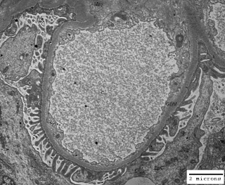
Just think of the life without space and you would understand its importance. Our body is like a tube open from both ends. You may appreciate space in your mouth (oral cavity), throat, nostrils, ears and lungs. In addition to these gross pockets of space there are hollow organs like heart (four chambers are there for blood flow regulation), gall bladder, urinary bladder and uterus (in females). Other examples of space within our body are cranial cavity, visceral cavity and cavities around all vital organs. There are micro-spaces in glandular tissues, alveoli of lungs, blood vessels and nephrons (glomeruli have capillary lumen and urinary space) in kidneys. In some of the renal disorders there are ultrastructural alterations in the areas/volumes of these micro-spaces within the kidneys leading to altered renal physiology and renal function. The figure-1 below illustrates normal urinary space (US) and capillary lumen (CL) or capillary space in a normal kidney; and figure-2 illustrates congestion of capillary lumen (CL) due to deposition of subendothelial deposits (SeD) in a kidney affected by lupus nephritis.

Figure-1: Ultramicrograph of a capillary loop from a normal human kidney illustrating normal urinary space (US) and capillary lumen (CL) with normal thickening of glomerular basement membrane (GBM); Uranyl acetate and Lead citrate stain.

Figure-2: Ultramicrograph of a capillary loop from human kidney affected by lupus nephritis, illustrating congestion of capillary lumen (CL) due to deposition of subendothelial deposits (SeD) with normal urinary space (US) but irregular thickening of glomerular basement membrane GBM); Uranyl acetate and Lead citrate stain.

In the illustration cited above you have seen the alteration in the space within the renal glomerulus. Abdominal tumors, brain tumors, polyps in the uterus, enlargement of spleen and liver, all these lead to functional as well as physiological changes in the body of a patient due to impact on space within the body.

Thursday, June 18, 2009

Therapy and Management of Glomerulonephritis

In my other articles you must have got ample information regarding classes or types of glomerulonephritis (GN). Primary glomerulonephritis accounts for about 30% patients requiring dialysis (a medical procedure of purifying blood with by passing through artificial kidney) and hospitalization. The yearly prevalence of primary GN is 0.002% that means 2 new patients per 100,000 population in a year. There are a variety of causes of primary GN with identical histopathological features. It is worth to mention here that there may be many immunopathological reasons in membranous glomerulonephritis (MGN). The character and severity of GN varies with the status of altered immune status of an individual. The understanding of pathogenesis of GN is must before initiating any therapy.

Regular follow-up in a clinic/renal clinic is must for the patient diagnosed of having a renal disease or glomerulonephritis. The follow-up provides an opportunity to the patient to learn about the complications of persistent GN and/or chronic renal failure. It has been observed that hypertension often develops coincidental with progression of renal disease. The hypertension needs to be kept under control in patients affected by persistent GN. The renal function deterioration causes edema in patients with glomerulonephritis. Though restriction in salt and water intake may cure the edema to some extent but diuretic drugs are preferred to treat the edema. Dietary management of progression of renal disease demands moderate reduction in protein intake (recommended: 0.8-1.0 g/kg body weight/day, during edema) to reduce the nephrotic overload and correction of proteinuria. Effective measures to reduce proteinuria are must to speed-up healing of renal lesions.

Corticosteroids, cyclophosphamide, chlorambucil and cyclosporin are the drugs of choice for the treatment of GN. About 95% of children generally respond to the first course of steroids within 8 weeks of commencement of treatment, whereas in adults it may take up-to 16 weeks and the percentage responding to the therapy could be around 80%. Oral prednisolone in single daily dose of 1mg/kg body weight is generally administered in adults. In children somewhat higher dose is required with reference to their body weights. Treatment of glomerulonephritis should never be tried as self help protocol as it needs regular follow-up. Parameters like body weight, blood pressure, 24 hour urinary protein, blood cells' count, blood urea and creatinine need to be worked out periodically to taper down the dose of steroids. Remission can also be achieved with cyclophosphamide, chlorambucil, cyclosporin and azathioprine. About 20-25% of patients are permanently cured with single course of treatment and around 50% may have relapse and need a repeat course of steroid treatment in combination with other immunosuppressive drugs. The dietary advice of nephrologist, controlled blood pressure and a treatment regimen for a sufficient time period may help a patient to keep a check on the complications of glomerulonephritis.

Saturday, May 30, 2009

Nephrotic Syndrome and Associated Renal Lesions

Nephrotic syndrome may occur in any type of primary or secondary glomerulonephritis. Around two dozen histopathological categories or subcategories of glomerulonephritis are now recognized and etiological factors are largely determined. However, prevention and treatment of glomerulonephritis needs momentum to curb the development of irreversible renal failure. The main diagnostic feature of nephrotic syndrome is massive proteinuria (excretion of protein in urine) exceeding 3 g/24 hour. The other features of nephrotic syndrome, such as, hypoproteinemia (decreased level of proteins in blood), edema (swelling) and hyperlipidemia (elevated levels of lipids in blood) are consequential due to excretion of proteins in urine. Clinically the patient does not bother to consult a nephrologist or general physician until edema becomes evident. With the fall in the plasma osmotic pressure due to loss of the plasma proteins in urine the fluid from blood would leak into the interstitial space resulting in a reduction in circulating blood volume, but the kidneys try to maintain blood volume by retaining salts and water.

In children around 80% cases of nephrotic syndrome are due to minimal change disease(MCD) and in adults the dominance of MCD is lost. In adults the cause of nephrotic syndrome may be minimal change disease, membranous glomerulonephritis (MGN), focal glomerulosclerosis, mesangial proliferative glomerulonephritis or membranoproliferative glomerulonephritis (MPGN). Metabolic disorders like diabetes mellitus could also be a cause of nephrotic syndrome. Secondary amyloidosis is also known to cause renal lesions associated with nephrotic syndrome. Immunological disorders like systemic lupus erythematosis and vasculitis may also be a cause of glomerulonephritis. Renal biopsy evaluation by light microscopy (LM), immunofluorescence microscopy (IFM) and electron microscopy (EM) is must for an accurate diagnosis of type of glomerulonephritis in a patient of nephrotic syndrome. Minimal change disease, membranous glomerulonephritis (MGN), focal glomerulosclerosis, mesangial proliferative glomerulonephritis, membranoproliferative glomerulonephritis (MPGN) and diffuse endocapillary glomerulonephritis are the common and primary causes of nephrotic syndrome. Focal segmental proliferative glomerulonephritis and diffuse proliferative glomerulonephritis with crescents are considered as uncommon-primary causes of nephrotic syndrome. Glomerulonephritis due to metabolic disorders, immunological disorders, toxemia of pregnancy or malignant conditions of kidney are labeled as secondary causes of nephrotic syndrome.

Friday, May 29, 2009

Bacterial Endocarditis and Associated Kidney Disease

There is very strong association between bacterial endocarditis or heart valve infection and kidney disease. Prior to the discovery, development and active use of antibiotics, the majority of the patients developed renal disease (kidney disease) as a consequence of subacute valvular infection. The incidence of clinical renal involvement has dropped significantly with the introduction of effective treatment of bacterial endocarditis with antibiotics. The assessment of renal involvement in bacterial endocarditis is quite difficult as transient changes in urine sediment are generally observed. There may be focal and segmental lesions with normal creatinine clearance. Intravenous drug users are at greater risk of developing bacterial endocarditis.

Staphylococcus aureus infection as the cause of bacterial endocarditis has been reported in majority of the cases that lead to a higher frequency of diffuse glomerular disease. The renal lesions associated with endocarditis involved embolization and infection. The renal lesions could also have immunological basis as immune complex deposits within glomeruli have been detected in majority of the cases. The involvement of complement (an immune response modulator protein in our blood) system during active disease (bacterial endocarditis) in association with immune complexes complicates the severity of intra-glomerular lesions. The two major categories of renal lesions found in patients affected by bacterial endocarditis are: (1) Focal segmental abnormality due to subacute infection and (2) Diffuse glomerular lesions in the patients with acute bacterial endocarditis mimicking the pattern of post-streptococcal glomerulonephritis. The electron micrograph (Fig-1) from the kidney biopsy of a patient with acute glomerulonephritis and acute bacterial endocarditis illustrates the subepithelial immune complex deposits.

Fig-1: Electron micrograph illustrating the hump shaped subepithelial immune complex deposits (D) alongside the glomerular basement membrane (GBM) and urinary space (US), during acute glomerulonephritis. Uranyl acetate and Lead citrate stain.

The assessment of renal involvement in endocarditis may be difficult diagnostic entity as only minor and transient changes are observed in urinary deposit with variable changes in blood biochemistry. The clinician must recognize the status of impaired cardiac output in the first stage and later workout the potential risk of treatment associated antibiotic nephrotoxicity. The assessment of renal function at the time of presentation of case could be helpful to rule out endocarditis associate renal disease or treatment associated antibiotic nephrotoxicity.

Thursday, April 9, 2009

Kidney Diseases and Elevated Levels of Blood Urea, Uric Acid and Creatinine

There could be minimal to gross impairment of renal function during the onset and progression of a kidney disease or renal disorder. This impairment of renal function may range from subclinical to complete renal failure. Urine analysis and blood biochemistry have been of great help in the assessment of renal function. Simultaneous increase in the levels of blood urea and uric acid has been observed during a variety of renal disorders. Uric acid is an end product of purine (a component of nucleic acids and nucleoproteins) metabolism. The level of uric acid in the blood depends on its endogenous production through purine metabolism as well as from the exogenously taken purines in the food items. Normal range of uric acid in blood is 2 - 6mg/dl. Elevated level of uric acid is also observed in gout. Urea is an end product of protein metabolism and its normal range in blood is 20 - 40mg/dl.

Formation of Urea: The amino acids derived by the digestion of proteins of the food we eat are absorbed by the villi of the small intestine and brought to the liver through the portal vein. The essential amino acids required for the growth and repair of body tissues are passed on to the blood circulation by the liver and others are used to produce the blood proteins and useful proteins for the body. Useless proteins are broken down in the liver to form bioenergy composed of carbon, hydrogen and oxygen and a waste product urea. Urea is a water soluble substance and carried away buy the blood stream.

The uric acid level may increase earlier than the blood urea level during the course of renal disease. The serum uric acid could be found markedly increased from the normal level of 2 -6mg/dl to 10 - 30mg/dl with minimal impairment of renal function. The creatinine level in blood starts rising after 2 to 4 fold rise in the blood urea level. The level of urea may rise in a variety of conditions, but increased level of creatinine is considered more severe than the increased level of blood urea. The creatinine is derived from the creatine and is a waste product; on the other hand the creatine is necessary for the muscle contraction and is related to the phosphocreatine breakdown. The normal level of creatinine in the blood plasma or serum is 1 - 2mg/dl and its normal daily excretion ranges from 1 to 2 grams. The serum creatinine values of up to and even exceeding occasionally 20mg/dl have been seen in the later stages of renal failure. The major cause of increased levels of serum creatinine and blood urea is the poor clearance of these substances by the kidneys rather than excessive production. In acute glomerulonephritis values from normal to over 300mg/dl are generally observed. In conditions such as malignant hypertension, chronic pyelonephritis and heavy metal poisoning 10 to 15 fold increase in blood urea level may be detected. However, in cases of hypoadrenalism (Addison's disease) blood urea level of about 100mg/dl could be detected. Fifteen to 20 fold increase in the level of blood urea (i.e. a level of 600 - 800mg/dl) may lead to uremic coma in more than 80% cases of cases affected by severe renal disease or renal failure.

Tuesday, April 7, 2009

Role of Adrenal Glands in Renal Physiology

The role of adrenal glands in the control of functioning of kidneys is very well established in terms of renal physiology. Every organ of our body has an embryonal origin and relationship with other organs. This relationship helps us to establish the physiology of systems associated with each other. There exists a pair of adrenal glands in our body and their position is suprarenal. Each adrenal gland has a cortex and medulla. There exists three layers of specialized cells in the adrenal cortex and these are: (1) Zona glomerulosa, (2) Zona fasciculata and (3) Zona reticularis. The medullae of adrenal glands belong to chromaffin system. The pituitary adrenocorticotrophic hormone (ACTH) controls the activity of adrenal glands in the production of cortisol (hydrocortisone). The inner medullary portion of the adrenal glands produces adrenaline (epinephrine) and noradrenaline (norepinephrine). The adrenal glands function under the control of sympathetic nervous system. The production of adrenal hormones increases in the conditions of emotions like anger or fear and states of asphyxia (lack of oxygen) and starvation and in turn raises the blood pressure in order to overcome the shock. The adrenaline epinephrine) influences the flow of urine and helps in the water balance by kidneys. Noradrenaline norepinephrine) is known to stimulate the muscle fibres in the walls of blood vessels, causing them to contract and thus raising the blood pressure. Adrenaline (epinephrine) also accelerates the carbohydrate metabolism by increasing the output of glucose from the liver.

The cortical layers of adrenal glands produce three types of steroids: (1) Mineralocorticoids are produced by the zona glomerulosa layer of the adrenal cortex and are associated with renal control of water and electrolytes. The mineralocorticoid naturally produced by the adrenal glands is aldosterone which controls the reabsorption of salts by the renal tubules. When there is deficiency of this hormone, too much water and sodium are lost from the body in the urine and too little potassium is excreted and this may lead to toxic levels of potassium in the blood. This results in polyuria, the passage of an excessive quantity of urine. The first of the adrenal steroids isolated or synthesized was the mineralocorticoid called desoxycorticosterone (DOC), which is used in the treatment of shock and polyuria, (2) Glucocorticoids are produced by the middle layer or zona fasciculata of the adrenal cortex and are associated with the metabolism of carbohydrates, fats and proteins. Glucocorticoids promote the conversion of proteins into glucose and storage of glucose as glycogen in the liver. Mineralocorticoids and glucocorticoids complement the action of each other and (3) Androsterone or androgens (sex hormones) are produced by the innermost layer or zona reticularis of the adrenal cortex. Androgens possess 19 carbon atoms in their chemical structure, with one oxygen atom attached to the 17th carbon atom and are also known as 17-ketosteroids and could normally be detected in the urine. Corticoids are chemically similar to cholesterol, sex hormones or corticosteroids. Our kidneys regulate the volume and composition of our body fluids in terms of water and electrolytes' balance through filtration, secretion, reabsorption and excretion. It is well established that kidneys function under the intelligent control of hormones, without which it would not be possible for the kidneys to maintain a state of homoeostasis in the body.

Monday, March 30, 2009

Kidney Diseases: Diagnostic Terms and Features

The genetic, environmental, chemical and biological factors are known to influence the bio-physiology and microanatomy of kidneys. A possible clinical diagnosis of kidney diseases or renal disorders could be achieved through ultrasonography, biochemical investigations of blood and urine analysis. The pathological diagnosis of non-neoplastic and neoplastic kidney diseases needs light microscopy (LM), immunofluorescence microscopy (IFM) and electron microscopy (EM) study of the kidney biopsy. Narrowing down at the appropriate and accurate diagnosis of a kidney disease needs expertise in the evaluation of LM, IFM and EM features. The light microcopy has its limitations in the exploration of microanatomy of renal lesions due to its low resolution power. The initial task in the pathological diagnosis of a kidney disease is to decide the renal compartments associated with the primary lesion or initial site of injury. The glomeruli, tubules, interstitium, extraglomerular vessels or podocytes may be affected primarily in various combinations in various renal diseases. The history of hypertension or diabetes in addition to chronic inflammatory disease like rheumatoid arthritis, osteomyelitis, tonsillitis and tuberculosis has its own implications in renal disorders. In some kidney diseases multiple components may be affected simultaneously by the pathogenic process. The glomeruli and blood vessels are found affected in certain forms of vasculitis. Immunological findings are mandatory to achieve an accurate diagnosis of vasculitis associated kidney diseases. Tubules and interstitium are found affected in tubulointerstitial nephritis. The role of EM and ultrastructural morphometry is implicit in achieving a diagnosis of thin basement membrane disease (TBMD), Alport's syndrome (hereditary nephropathy), minimal change disease (MCD), amyloidosis and evaluation of podocyte injury. The thickness and texture of glomerular basement membrane (GBM), reorganization of foot processes of podocytes and podocyte injury are directly associated with the biophysiology of proteinuria (excretion of protein in urine) and hematuria in some kidney diseases. The histopathologic lesions in the affected kidneys could only be explained with a thorough knowledge of universally accepted appropriate terms which could be understood by a clinician. The term focal is used when <50% of glomeruli are involved and the term diffuse refers to the involvement of 50% or more glomeruli. The term segmental is used when a part of a glomerular tuft is affected and the term global is used when entire glomerular tuft is affected. The term mesangial hypercellularity means >4 nuclei in the matrix of a peripheral mesangial segment. The term sclerosis refers to increased collagenous extracellular matrix causing mesangial expansion, obliterating capillary lumen or forming contact to Bowman's capsule. Some of the important diagnostic features of kidney biopsy evaluation have been cited below in a tabulated form: (For a full view of the Table - Just click on the image below)

The neoplastic kidney disease are renal cell carcinoma, juxta glomerular cell tumor, renal adenoma, oncocytoma and metastatic tumors which need immunohistochemical (IHC) and EM study for an accurate diagnosis.

Saturday, March 28, 2009

Urinary Deposits in Health and Disease

The water and salt balance of our body is taken care by our kidneys through excretion of water and salts under the strict regulatory control of various hormones. The chemical and microscopic examination of urine for the evaluation of health status is a routine procedure at health centers. The abnormal excretion of biochemical substances on physical and chemical analysis of urine and presence of chemical crystals, various cell types and casts in the urine is the first alarming point about many diseases. The microscopic examination of urinary deposits would yield a valuable information about a positive or negative character. The components of the urinary deposit can be classified into three groups: 1) Chemicals as crystals or amorphous deposits, 2) Cells from the blood or urinary tract, and 3) Casts

  1. Chemical substances as crystals or amorphous deposits: Some of the inorganic and organic chemical substances could be appreciated in the urine of normal people of all age groups, but some other chemicals are always associated with pathological conditions. The presence of crystals of chemical substances in the urinary deposits is influenced by acidic or alkaline reaction of the urine. Phosphates (ammonium magnesium phosphate, Calcium hydrogen phosphate and magnesium phosphate), Calcium oxalate, uric acid and urates (of Ammonium, Sodium, Potassium, Calcium and Magnesium) are most commonly detected in the urinary deposits. Other chemical substances viewed in the urinary deposits may be Calcium carbonate, Calcium sulphate, amino acids (cystine, tyrosine and leucine), hippuric acid, cholesterol, xanthine, Sulphonamide drugs and pigments like bilirubin. Phosphates are deposited in alkaline urine and get dissolved in dilute acetic acid. Calcium oxalate crystals are soluble in hydrochloric acid but uric acid crystals are not soluble in acetic acid or hydrochloric acid. Presence of red blood cells(RBCs) in the urine may give color to the deposits. The crystals of chemical substance have very typical shapes. Microscopic examination of urinary deposits by an experienced medical technologist or pathologist is needed for an accurate assessment of chemicals, cells or casts excreted in the urine. Calcium oxalate is present in some fruits and vegetables and notable among them are strawberries, rhubarb and spinach. The crystals of Tyrosine appear like tufts of needles and those of Leucine are in spherical shape. Crystals of tyrosine and leucine are seen very rarely in the cases of severe liver disease and cirrhosis. Crystals of cholesterol appear as rectangular or rhomboid plates with notched corners and occur the urine form the patients affected by some kidney disease. Sulphonamide crystals could be found in urinary deposits during treatment with such drugs. These are formed from acetyl derivatives in the urinary tract.
  2. Cells: Red blood cells (RBCs) could be detected in the urinary deposits during macrohematuria (>1000 RBCs/ml of urine) as well as microhematuria (<1000 RBCs/ml of urine). Pus cells: Less than 10 leucocytes or pus cells per microlitre (µl) of urine may occur in normal urine. An increase in the number of pus cells is called pyuria and is indicative of some inflammatory disease in the urinary tract. Urine culture may help to establish the causative agent of urinary tract infection. The cells present during the acute inflammation are mainly polymorphonuclear cells. Microscopic examination of the urinary deposit is the only satisfactory test to establish the presence of pus cells. Epithelial Cells: Epithelial cells may be detected normally in urine from female patients but an increased number could be due to pathological reasons. Epithelial cells in the urine of males are normally very few in number. Epithelial cells could be from the squamous epithelium, transitional epithelium (from the bladder, prostate, ureters and pelvis of kidneys), and basal or parabasal cells. Abnormal cells such as tumor cells may also be detected in the urinary deposits. Sometimes spermatozoa may also be present in the normal urine of males.
  3. Casts: Casts are formed in renal tubules whose shape these take. Subsequently the casts are pushed by the fluid along the tubules and appear in the urine. These are seen on microscopic examination of urinary deposits. The casts could be classified on the basis of their appearance under the microscope as: 1) Hyaline casts: are simplest, pale transparent and homogenous structures with cylindrical shape and do not contain cells or granules. 2) Epithelial casts: When there is tubular damage, cells from the tubular epithelium could be trapped into casts and give rise to epithelial casts. 3) Granular casts: Casts containing closely packed granules of various size and shape are generally formed due to degeneration of tubular epithelial cells and are indicative of renal disease. 4) Fatty casts: are derived from epithelial cells when fat granules are present along with granular material. Such casts are found in tubulopathy and are indicative of degeneration of tubular epithelium.

Important Points:

  • Collection of urine from each kidney by ureteric catheterization and from urinary bladder may be used to locate the site of pus formation.
  • Cytological examination of the first morning urine for three consecutive days should be performed to rule out any malignancy in doubtful case.
  • If large number of pus cells are detected in the urine, a urine culture is advisable to rule out the infectious organism.

Tuesday, March 24, 2009

Types and Causes of Proteinuria

Proteinuria means the excretion of protein in the urine. A healthy person does not excrete proteins in the urine or the excretion of proteins is less than 150 mg per day. The proteins most commonly found in the urine are those derived from the plasma of blood and consist of a mixture of albumin and globulin. Predominantly albuminuria (excretion of albumin in urine) is detectable on routine urine analysis during a medical examination. Albuminuria could be organic (due to involvement of kidneys or other organs) or functional (due to physiological or biological stress on kidneys). The functional albuminuria is usually intermittent and not accompanied by any symptoms or evidence of kidney disease. Renal function tests and urinary deposits are found to be normal during the functional albuminuria. It may be connected with posture; being absent when the person is lying down and present when standing. The functional albuminuria usually clears up in early adult life and seems to be associated with the growth and development of kidneys. Any severe stress may also lead to transient albuminuria. Exposure to severe cold and excessive exercise or physical activity may cause functional or transient proteinuria. However, there is nothing to worry about as the functional albuminuria is self limiting with respect to the cause. Mild to moderate functional albuminuria may also be detected during last two months of pregnancy due to pressure on kidneys.

Organic albuminuria is of three types: 1) Renal Albuminuria - When the cause is the kidney disease. 2) Pre-renal Albuminuria - When the kidneys are affected secondarily to some other disease. Post-renal Albuminuria - When the protein is added to the urine after it has left the renal tubules.

  1. Renal Albuminuria: It is found in all forms of kidney disease. The cause of renal disorder or kidney disease may be inflammatory (infectious), degenerative (immunological) or destructive (toxic or malignant). The plasma globulin and red blood cells (RBCs) may also be excreted along with albumin during some renal disorders. The urine would be smoky in color if macroscopic hematuria (blood in urine) is also associated with proteinuria. The cases of acute glomerulonephritis may excrete 0.5 to 2.0 percent (0.5 g to 2.0 g/dl) protein in the urine, whereas the cases affected by chronic glomerulonephritis generally excrete less than 0.5 percent (0.5 g/dl) protein in the urine. The amount of protein excreted daily would vary depending on the volume of urine voided daily. The ratio of albumin to globulin excreted in the urine may vary from 10:1 to 5:1. A routine and quantitative urine analysis is required to evaluate the extent of excretion of proteins in the urine.

  2. Pre-renal Albuminuria: It is found in a variety of conditions exerting stress on the kidneys. The pre-renal albuminuria usually disappears when the primary disease is cured. Impairment of renal circulation due to dehydration, diarrhea or vomiting, blood loss due to accidental injuries or anemia are the most common conditions, which could lead to pre-renal albuminuria.

  3. Post-renal Albuminuria: The proteinuria or albuminuria is termed as post-renal albuminuria if protein is possibly added to the urine as it passes along the urinary tract after leaving the urinary tubules of the kidneys. The major causes of the post-renal albuminuria are the lesions of the renal pelvis or urinary bladder. Lesions of the prostate (in male patients) and urethra also lead to post-renal albuminuria. Admixture of discharges from the vagina (in female patients) and semen (in male patients) may also give positive tests for protein.

Wednesday, February 11, 2009

Micturition Disorders and Neurogenic Bladder

Micturition:

The process of voiding urine is called micturition. The urine is formed in the kidneys and collected into the urinary bladder. The urinary bladder is connected to kidneys through a pair of ureters. When a pressure of accumulated urine develops in the urinary bladder, we have a desire to urinate. An accumulation of 150 ml to 200 ml of urine activates the nerve endings in the muscular wall of the urinary bladder. The micturition is a reflex act controlled and inhibited by the higher centers in our brain. The act leads to the contraction of the muscular coat of the urinary bladder and relaxation of sphincter muscles. The urinary bladder is controlled by the pelvic nerves and the synaptic fibers from the hypogastric plexus. It may be voluntarily assisted by the contraction of abdominal muscles by increasing pressure in the abdominal cavity. The voluntary contraction of abdominal muscles exerts pressure on the visceral organs and in turn on the urinary bladder and helps in emptying of the bladder.

Neurogenic Bladder:

As stated above, the urinary bladder activity is controlled by the pelvic nerves and the sympathetic nerve fibers from the hypogastric plexus. A condition may arise due to a variety of causes leading to an interruption of the nerve messages between the brain and the urinary bladder. Due to lack of dynamic control over the muscles of the urinary bladder, the bladder fails to store or release the urine properly. Spinal cord injury due to some fatal accident, brain tumor, stroke, diseases such as multiple sclerosis or diabetes mellitus and congenital disorders could be the cause of neurogenic bladder.

There may be varied symptoms of neurogenic bladder depending on the lesion and the site of the injury and severity of injury. The patient may express inability to store the urine, excessive frequency of micturition and incontinence. All this is caused by overactive urinary bladder or a weak sphincter (outlet controlling muscles). On the other hand a weak urinary bladder or an over tight sphincter may lead to retention of urine and difficulty in urinating. Though the problem is associated with the urinary system but the cause is neurological and only a neurologist could help the patient in the right perspective. Renal function tests and ultrasound examination is must to evaluate the status of kidneys. A thorough evaluation is needed to ascertain the cause of neurogenic bladder disorder. Urodynamic testing with video x-rays and uroflometry should be carried out to evaluate the extent of emptying of the urinary bladder.

Monday, February 9, 2009

Altered Physiology and Diseases of the Muscles

The skeletal muscles or flesh are essential for the motor functions as well as the shape and features of our body. The muscular atrophy (shrinkage of muscle mass) may lead to a variety of diseases of the muscles. These diseases are distinguished with respect o the site of origin of a disorder as mentioned below:

1. Origin in motor neurons: Polymyelitis, progressive muscular atrophy

2. Origin in the nerve fibers: Polyneuritis

3. Origin in the myoneural junction: Myasthenia gravis, myotonia congenital, and

4. Origin in the muscle only: Primary muscular dystrophy.

All voluntary muscle have innervation by motor neurons. The muscle fibers are composed of myofibrils and these fibrils appear to be the ultimate functional units of the muscle and have alternate light and dark bands. The contractile protein of the muscle is called actomycin (AM) and is composed of actin and myosin. The actomycin is considered to be the structural protein of the resting myofibrils and contain many enzymes necessary the muscle metabolism. The organic phosphate compound called adenosine triphosphate (ATP) is an energy rich compound and is attached to actomycin of myofibrils. The neuronal spark of the motor neurons fires the adenosine triphosphate-actomycin linkage and breaks it. The adenosine triphosphate (ATP) is liberated and its breakdown provides energy for the muscle contraction. During contraction of the muscle, the actin threads could be shortened to 40% of its original relaxed phase size. The physiology of muscle contraction involves a series of molecular interactions and rearrangements within the complex structure of actin, myosin, and adenosine triphosphate.

The change in the functions of any part of the above cited metabolic pathway may lead muscle weakness. The microscopic examination of muscle biopsy may help to achieve a proper diagnosis. There muscle biopsy could reveal an involvement of a whole bundle of muscle fibers in the cases with neurogenic origin. I cases affected by primary muscular dystrophy only some the fibrils may be involved. The ultrastructural study of the muscle biopsy by a transmission electron microscope is always helpful in achieving an accurate diagnosis of the diseases of the muscles.

Physiology of Muscle Contraction

The essential function of the skeletal muscle is contraction and relaxation in response to a command from a motor neuron. The muscle is very unique organ and is capable converting stored chemical energy into mechanical energy through metabolic processes. The skeletal muscles constitute from 40 to 45 percent of the body weight in an adult. There are 434 voluntary muscles in our body and about 250 million muscle fibers constitute these muscles. The muscle cells or sarcomeres are very complex in structure, metabolism and functions.

All voluntary muscle have innervation by motor neurons. The muscle fibers are composed of myofibrils within the sarcoplasm. The myofibrils appear to be the ultimate functional units of the muscle and have alternate light and dark bands. The contractile protein of the muscle is called actomycin (AM) and is composed of two components: (1) actin and (2) myosin. The actomycin is considered to be the structural protein of the resting myofibrils and contain many enzymes necessary the muscle metabolism. The organic phosphate compound called adenosine triphosphate (ATP) is an energy rich compound and is attached to actomycin of myofibrils. The neuronal spark of the motor neurons fires the ATP-AM linkage and breaks it. The ATP is liberated and its breakdown provides energy for the muscle contraction. During contraction of the muscle, the actin threads could be shortened to 40% of its original relaxed phase size. Physiology of muscle contraction involves a series of molecular interactions and rearrangements within the system composed of actin, myosin, and adenosine triphosphate.

Friday, February 6, 2009

Role of Grey Matter and White Matter in Our Nervous System

Our nervous system could be studied under two main headings for quick understanding: (1) The Central Nervous System (CNS) or Cerebrospinal Nervous System and (2) The Autonomic Nervous System. The autonomic nervous system includes the sympathetic nervous system and parasympathetic nervous system.

The central nervous system includes the brain and the spinal cord, and the nerves arising out from these. The nerves arising from the brain and the spinal cord are called peripheral nerves. The nervous tissue is one of the four elementary tissues of our body and commands the sensory and motor functions of our body in addition to intellect, emotions and memory functions. The nervous tissue is composed of nerve cells, dendrites, nerve fibres and nerve endings. The composite mass of nerve cells is called grey matter. The grey matter is found in the cortex of the brain, inner part of the spinal cord and cerebral ganglia. The role of the grey matter is the processing of the sensory and motor information, control of emotions, memory and intellect. The control of emotions, memory and intellect is associated with the growth and development of the brain. The nerve fibres and or axons constitute the white matter. The white color of the sheath of fatty matter called the myelin sheath covering the axons or nerve fibres is the reason behind the nomenclature as white matter. The white matter is the major component of the central portion of cerebral part of brain. The myelin sheath serves to protect, nourish and insulate the nerve fibres from each other. A nerve cell along with its dendrites, nerve fibre or axon and nerve endings is called a neuron as depicted in the figure-1 below:

Figure-1: Components of a Neuron

There are two types of neurons in our body. The neurons which carry impulses out from the brain to the tissues are called efferent neurons and the neurons which carry impulses to the brain from the tissues are called afferent neurons. Efferent neurons which supply impulses to the muscles and produce movement are called motor neurons and which supply impulses to the glands and produce secretion are called secretory neurons. Afferent neurons are also known as sensory neurons since these help us to assess a feeling of touch, pain and heat or cold.

Tuesday, February 3, 2009

Quick Tips on the Surface Anatomy of Our Body

Our own body is a live specimen for understanding the surface anatomy of human body. Knowledge of surface anatomy may help us to understand the diagnostic terms used by a physician or surgeon. The physical examination parameters of a patient are recorded with reference to certain landmark points which are universally understood. By inspection, palpitation, percussion and auscultations a physician or surgeon studies condition of his/her patient to arrive at a diagnosis. The position of many internal structures and organs is described in relation to various bony points termed as landmark points.

There are some land mark points of surface anatomy of the head; like longitudinal fissure, superior longitudinal sinus, central sucus or fissure of Ronaldo, mastoid processes ete. There are anterior and posterior triangles of neck formed by sternomastoid muscle. In the trunk many of the organs are being described in relation to their surface anatomy. The apex beat of the heart can be heard in the fifth intercostal space, 31/2 inches from the midline. The abdomen is divided into 9 parts by four imaginary lines (two vertical and two horizontal). These 9 parts have been depicted in the figure-1 below:

Figure-1: Abdominal Regions (1: Right Hypochondriac Region, 2: Epigastric Region, 3: Left Hypochondriac Region, 4: Right Lumbar Region, 5: Umbilical Region, 6: Left Lumbar Region, 7: Right Iliac Region, 8: Hypogastric, 9: Left Iliac Region.

The liver occupies parts of the right hypochondriac and epigastric areas, and extends transversely into the left hypochondriac region and also occupies a part of the right lumbar region. The lungs and the heart occupy the thoracic cavity and are well secured there in the protective bony cage. The spleen lies on the left side beneath the ninth, tenth and eleventh ribs. The left kidney lies between the eleventh thoracic to the third lumbar vertebra. The right kidney is slightly lower as its upper pole touches the lower part of the liver.

The rectus abdominus could be felt along each side of the middle line of the abdomen. The umbilicus lies on level with the disc between the third and fourth lumbar vertebrae. The stomach lies in the upper and left aspect of the abdomen, partly behind the lower ribs and cartilages. The fundus of the stomach reaches as high as the fifth left intercostal space. The gall bladder projects slightly from the costal margins at the level of the ninth right costal cartilage. The pancreas lies at the back of the abdominal cavity across the first lumbar vertebra. The aorta passes through the common iliac arteries in front of the fourth lumbar vertebra. The caecum is present on the right side and the commencement of the sigmoid fissure of the colon lie respectively in the right and left iliac fossae.

Friday, January 30, 2009

Why Respiration is Necessary for Propagation of Life ?

One can survive without food and water for many hours but survival without the air or oxygen is not possible beyond a few minutes. Every cell in our body is programmed to perform a definite metabolic function necessary for our life. By means of breathing we assimilate oxygen from the air for intracellular metabolism. Through metabolic processes, cells of our body release carbon dioxide (CO2) and water (H2O), which are transported away by the circulating blood. Respiration is a two fold process: (1) The interchange of gases in the lungs is called external respiration and (2) The interchange of gases in the tissues is called internal respiration. In the external respiration, oxygen is taken in through the nose and mouth and flows down to the alveoli in the lungs, where it comes in contact with the alveolar capillary membrane and is taken up by the hemoglobin of the red blood cells (RBCs). The oxygenated blood goes to the heart and is further pumped to the various parts of our body. In the lungs, carbon dioxide (CO2), a waste product of the metabolism is released out through the alveolar capillary membranes and breathed out through the nose and mouth.

The air we breathe in, contains 79% of nitrogen, 20% of oxygen and 0.04 carbon dioxide (CO2) along with atmospheric water vapors; where as the air we breathe out, contains 79% of nitrogen, 16% of oxygen and 4.04 carbon dioxide (CO2) along with water vapors released from the alveoli. The total air capacity of our lungs is about 4.5 to 5.0 liters of air and only 1/10th (500 ml) is generally inspired or expired. The volume of the air under exchange is also termed as tidal air. Our breathing is controlled by two factors: (1) The chemical control of respiration and (2) The nervous control of respiration through the medulla oblongata of our brain. The normal rate of respiration in different age groups is as under:

Status of AgeRespiration Rate per minute 
Newborn 38 - 42 
Up to 12 months 28 - 32 
2 years to 5 years 24 - 26 
6 years to 16 years 20 - 24 
Adults  10 - 20 

The oxygen need of our body varies with the type of activity we perform. Rate of respiration goes up during exercise or running and is considered a vital health parameter for medical fitness evaluation of an individual. As stated above the oxygen (O2) is the essence of the life force and lack of it may lead to a state of hypoxia or anoxia and brain damage.

Thursday, January 29, 2009

What causes Pyelonephritis and How Serious is It?

The pyelonephritis is said to be very serious renal disease. The well documented cause of pyelonephritis is an infection of interstitial (intertubular) tissue with pyogenic bacteria, most frequently E. coli and Staphylococci, and sometimes Proteus vulgaris and Pseudomonas pyocyaneus. In almost 66% of cases the infection could be secondary to the urinary tract infection and in 33% of cases the infection could be primary through infection of blood. The ascending infection of kidney or kidneys from the urinary tract is quite common in infants. There could be ascending infection of kidneys in pregnant women and also in women with the cancer of the cervix. In the elderly male patients, the ascending infection of kidneys is possible if their prostate is enlarged and the urethra is obstructed causing partial or full retention of urine. Irrespective of the sex of the patient, the obstruction plays a vital role in causing the infection of kidneys. It has been proved through experimental study on rabbits that the obstruction of urinary system was the main cause of renal infection and pyelonephritis. In man infection of blood may cause unilateral or bilateral pyelonephritis depending on the obstruction. The obstruction may be functional or organic. The ultrasonography or the serial radiographical study is required to rule out the obstruction.

The pyelonephritis in its chronic form is very dangerous disease. The kidney may become a bag of pus due to infection with pyogenic bacteria cited above. The condition is also called pyonephrosis. A pure hydronephrosis (nephrosis caused due to back pressure of accumulated urine) in an advanced stage may become infected. The kidneys get much enlarged due to hydronephrosis or pyelonephritis. There is possibility of complete erosion of functional architecture in side the kidney due to pyogenic infection. Pyelonephritis may also develop in a gradual manner with little frank suppuration. The early detection of disease could be better for the patient as it could be treated effectively.

Thursday, January 22, 2009

Acute Glomerulonephritis

The term acute glomerulonephritis is used by clinicians as well as pathologists to describe the sudden onset of kidney disease. Irrespective of the cause, there would be enlargement of a kidney or kidneys and the capsule around the kidney is strained and stretched. Ultrasonography is always helpful to ascertain the size of kidneys. Histological examination of kidney biopsy would present densely cellular glomerular tuft with polymorphonuclear cells (a type of white blood cells) in the glomerular capillaries. Accumulation of leukocytes (white blood cells) in the glomerular capillaries with swelling and proliferation of the vascular endothelium (inner lining of capillaries) is rapidly followed by edema (swelling) and mesangial proliferation (enlargement of inner stalk of a cluster of glomerular capillaries)leading to capillary ischemia (poor blood supply).

The glomerular basement membrane (GBM) is the other important component affected by acute glomerulonephritis. By electron microscopic study of kidney biopsy, electron dense deposits of antigen-antibody complexes could be revealed in and around the glomerular basement membrane. The ultrastructural features of normal GBM have been depicted in the figure-1 and the figure-2 is from a case affected by acute glomerulonephritis.

Figure-1: Electron micrograph of a capillary loop of glomerular tuft showing normal features; CL: capillary lumen, US; urinary space, En: endothelium, GBM: glomerular basement membrane, EpC: epithelial cell or podocyte.

Figure-2: Electron micrograph of a capillary loop of glomerular tuft from a case affected by acute glomerulonephritis is showing sub-epithelial deposits; CL: capillary lumen, US; urinary space, GBM: glomerular basement membrane, dep: deposits on sub-epithelial site of GBM.

The tubules in the kidney may show slight degenerative changes. The degree of degenerative changes in the tubules depends on the extent of the glomerular obstruction. The interstitial tissue and the blood vessels are observed to be normal on the histological study of kidney biopsy.

Saturday, January 10, 2009

The Mind and Brain: Functions and Physiology

The most important fact is that the brain is the organ of the mind and the mind is like the software of a computer. The mental functions are related to the cerebral cortex of the brain. In humans the cerebral cortex is better developed than the inferior animals. Our sense organs are like input devices of a computer. Our brain processes and stores the data in encapsulated form with respect to attributes. Our ears transmit sound signals to our brain, which are analyzed by our mind and classified as noise, vocal audio and music. Music files are further categorized as per specific attributes and stored in our brain. Whenever we listen to the same sound again we could easily name a person as per tonal quality of voice, instrument as per the pitch and rhythm of the music and the birds and animals as per their specific vocal attributes. Human mind is capable of distinguishing a variety of fragrances and odors, human faces and pictures, fonts and shapes, smooth and rough surfaces, bitter, sweet or sour tastes etc. Expressions of thoughts and emotions are special functions of our mind.

The brain, like other organs is powered by chemicals. Whenever the chemistry of brain is disturbed, mental symptoms are an early result. The glucose and oxygen have a vital role in the physiology and bio-energy generation of our body. The anoxia (low oxygen supply) of airmen and in the early stages of anesthesia may lead to chock. The hypoglycemia in diabetics on insulin therapy has been known to cause insulin shock or hypoglycemic shock. Certain groups of chemicals or drugs can cause mental disturbance and there are other chemicals or drugs which are capable of improving the mental function. For example hallucinogens like LSD disturb the mental functioning of normal people whereas tranquilizers and psychic energizers are known to improve the mental functioning of abnormal people. Except the numerical ability, the human mind and brain are faster than any computer in answering a variety of questions related to memory and experiences. Till date no computer could store the attributes of taste, smell, feelings and emotions. The super programming and coding of our mind is updated every moment through our observation and experience.

Thursday, January 8, 2009

Azotemic or Hydropic Glomerulonephritis

The high concentration of non protein nitrogen (NPN) in the blood of a patient, mainly due to elevated level of urea is termed as azotemia. Elevated level of urea in blood is termed as uremia and it may be due to renal abnormality or due to other health problems like dehydration and excessive burns on the body. The glomerulonephritis (inflammation of glomeruli of kidneys) may be azotemic or hydropic type depending on the nature of glomerular lesions. There may be marked narrowing of the glomerular capillaries leading to azotemia with renal insufficiency and hypertension. On the other hand the status of glomerular capillaries may remain normal but there could be an increase in the permeability of glomerular basement membrane (GBM), the filtration barrier of kidneys. The clinical picture would be hydropic (accumulation of water in the tissues of the body) in character. The hydropic glomerulonephritis is clinically represented with edema associated with hypoproteinemia (low level of protein in blood) and hyperlipemia (high concentration of lipids or cholesterol in blood).

A child or an adult affected with fever or some other acute disease finds the increase in the daily output of urine. There may be tinge of blood in the urine of the patient affected by glomerulonephritis. Sudden appearance of features like puffiness or swelling on face (facial edema), ankles and hands after any acute disease needs expert medical attention and medication. The urine of such patients would show notable excretion of albumin. The patient may be less perspiring with dry skin. The pulse could be full and hard. The loin pain and a feeling of heaviness in the lower abdominal region are the associated symptoms. The polyuria (excessive output of urine) is probably compensatory action of affected kidneys to flush out solid wastes of metabolism form the body. An effective therapy would definitely reverse the associated symptoms and lesions.