Tuesday, June 30, 2009

The Space Within and Outside Our Body

The space has a great role in our life. Our body is composed of five basic components: the earth, water, air, fire (heat or temperature) and the sky or space. The space within and outside our body is must for the existence of life. The outer space is composed of air (mixture of gases), vapours, finer particles, microorganisms, radiation, light, cold and heat. The composition of environment influences our breathing, metabolism and physiology. Our body reacts in a variety of ways to the external space and the environment possessed by it. In fact the particles floating in the air or transmitted through it may cause allergic reactions, infections, hot or cold skin burns or even skin cancer. All activities of human beings or animals are space oriented.

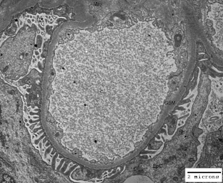
Just think of the life without space and you would understand its importance. Our body is like a tube open from both ends. You may appreciate space in your mouth (oral cavity), throat, nostrils, ears and lungs. In addition to these gross pockets of space there are hollow organs like heart (four chambers are there for blood flow regulation), gall bladder, urinary bladder and uterus (in females). Other examples of space within our body are cranial cavity, visceral cavity and cavities around all vital organs. There are micro-spaces in glandular tissues, alveoli of lungs, blood vessels and nephrons (glomeruli have capillary lumen and urinary space) in kidneys. In some of the renal disorders there are ultrastructural alterations in the areas/volumes of these micro-spaces within the kidneys leading to altered renal physiology and renal function. The figure-1 below illustrates normal urinary space (US) and capillary lumen (CL) or capillary space in a normal kidney; and figure-2 illustrates congestion of capillary lumen (CL) due to deposition of subendothelial deposits (SeD) in a kidney affected by lupus nephritis.

Figure-1: Ultramicrograph of a capillary loop from a normal human kidney illustrating normal urinary space (US) and capillary lumen (CL) with normal thickening of glomerular basement membrane (GBM); Uranyl acetate and Lead citrate stain.

Figure-2: Ultramicrograph of a capillary loop from human kidney affected by lupus nephritis, illustrating congestion of capillary lumen (CL) due to deposition of subendothelial deposits (SeD) with normal urinary space (US) but irregular thickening of glomerular basement membrane GBM); Uranyl acetate and Lead citrate stain.

In the illustration cited above you have seen the alteration in the space within the renal glomerulus. Abdominal tumors, brain tumors, polyps in the uterus, enlargement of spleen and liver, all these lead to functional as well as physiological changes in the body of a patient due to impact on space within the body.
Thursday, June 18, 2009

Therapy and Management of Glomerulonephritis

In my other articles you must have got ample information regarding classes or types of glomerulonephritis (GN). Primary glomerulonephritis accounts for about 30% patients requiring dialysis (a medical procedure of purifying blood with by passing through artificial kidney) and hospitalization. The yearly prevalence of primary GN is 0.002% that means 2 new patients per 100,000 population in a year. There are a variety of causes of primary GN with identical histopathological features. It is worth to mention here that there may be many immunopathological reasons in membranous glomerulonephritis (MGN). The character and severity of GN varies with the status of altered immune status of an individual. The understanding of pathogenesis of GN is must before initiating any therapy.

Regular follow-up in a clinic/renal clinic is must for the patient diagnosed of having a renal disease or glomerulonephritis. The follow-up provides an opportunity to the patient to learn about the complications of persistent GN and/or chronic renal failure. It has been observed that hypertension often develops coincidental with progression of renal disease. The hypertension needs to be kept under control in patients affected by persistent GN. The renal function deterioration causes edema in patients with glomerulonephritis. Though restriction in salt and water intake may cure the edema to some extent but diuretic drugs are preferred to treat the edema. Dietary management of progression of renal disease demands moderate reduction in protein intake (recommended: 0.8-1.0 g/kg body weight/day, during edema) to reduce the nephrotic overload and correction of proteinuria. Effective measures to reduce proteinuria are must to speed-up healing of renal lesions.

Corticosteroids, cyclophosphamide, chlorambucil and cyclosporin are the drugs of choice for the treatment of GN. About 95% of children generally respond to the first course of steroids within 8 weeks of commencement of treatment, whereas in adults it may take up-to 16 weeks and the percentage responding to the therapy could be around 80%. Oral prednisolone in single daily dose of 1mg/kg body weight is generally administered in adults. In children somewhat higher dose is required with reference to their body weights. Treatment of glomerulonephritis should never be tried as self help protocol as it needs regular follow-up. Parameters like body weight, blood pressure, 24 hour urinary protein, blood cells' count, blood urea and creatinine need to be worked out periodically to taper down the dose of steroids. Remission can also be achieved with cyclophosphamide, chlorambucil, cyclosporin and azathioprine. About 20-25% of patients are permanently cured with single course of treatment and around 50% may have relapse and need a repeat course of steroid treatment in combination with other immunosuppressive drugs. The dietary advice of nephrologist, controlled blood pressure and a treatment regimen for a sufficient time period may help a patient to keep a check on the complications of glomerulonephritis.