Showing posts with label glomeruli. Show all posts

Tuesday, July 27, 2010

Podocytes and Podocytopathies

Kidneys play a vital role in excretion and water/fluid volume regulation. Glomeruli are the filtration units of nephrons in the kidneys and these contain cellular and non cellular components in addition to capillary space and urinary space. Podocytes (cells with pedicles or feet) are post-mitotic epithelial cells resting in the urinary space of glomeruli. The number, size and morphology of podocytes are influenced by biochemical, immunological, therapeutic and genetic factors. According to the old classification of renal disorders, the patients having nephrotic syndrome can be grouped into two groups: (1) Non-immune complex mediated nephrotic syndrome, and (2) Immune complex mediated nephrotic syndrome. Now, patients with non-immune complex mediated nephrotic syndrome may have three possible diagnoses:

  1. Minimal change disease: Wherein morphologic evaluation of the renal biopsy (kidney biopsy) by light microscopy does not exhibit any glomerular damage. However, extensive effacement of foot processes of podocytes can be revealed by electron microscopy.
  2. Focal segmental glomerulosclerosis (FSGS): Wherein segmental sclerosis/solidification of the glomerular tuft, along with hyalinosis and adhesion of tuft to the Bowman's capsule is exhibited on the renal biopsy (kidney biopsy) evaluation by light microscopy. In these cases, variable degree of foot process effacement can be revealed by electron microscopy.
  3. Collapsing glomerulopathy: FSGS associated with the rapid deterioration of renal function was described as "Malignant FSGS" in 1978. During HIV pandemic in 1980's the associated nephropathy showing collapse of glomerular capillary wall along with increased cellularity in the urinary space was termed as HIV associated nephropathy (HIV-AN). Collapsing glomerulopathy was first time described in non-HIV patients in 1986 by Weiss and associates.

Now we know that podocyte number and effacement of their foot processes due to genetic or biological factors are very much associated with the primary nephrotic syndrome or proteinuric renal disorders. The etiology and pathogenic mechanisms are known to influence the morphologic diagnosis of podocytopathies. Podocytopathies are proteinuric renal disorders caused due to intrinsic or extrinsic podocyte injury exhibited by variable degree of foot process effacement and altered genotypic and/or phenotypic expression. Podocytes may reorganize their foot processes (altered cell morphology without change in cell count/number). There may be decreased number of podocytes (podocytopenia) if the injured podocytes die. There may be podocyte developmental arrest as seen in congenital nephrotic syndrome of Finnish type (CNF). Podocytes may dedifferentiate and proliferate under genetic, immunological, viral or therapeutic insult and re-enter the cell cycle despite the fact that podocytes are post-mitotic cells. Two electron micrographs are exhibited below to illustrate the normal (Figure-1) and increased number(Figure-2) podocytes in the urinary space of glomeruli from different cases.

Figure-1: Electron micrograph through a portion of glomerulus from a case of minimal change disease showing normal number of podocytes. (GBM: glomerular basement membrane, CL: capillary lumen, EnC: Endothelial cell, US: urinary space and Pc: podocyte)

Figure-2: Electron micrograph through a portion of glomerulus from a case of podocytopathy showing increased number of podocytes. (GBM: glomerular basement membrane, CL: capillary lumen, US: urinary space and Pc: podocytes)

Monday, September 28, 2009

IgA Nephropathy as a cause of End Stage Renal Disease

There are a variety of causes of end stage renal disease (ESRD) in teenagers and adults. Immunoglobulin-A (IgA) nephropathy could be a cause of end stage renal disease (ESRD) in around 25% of cases. There are five types of immunoglobulins in our body for protection against microorganisms and IgA provides defence at mucous membranes. Colostrum and breast milk are rich sources of IgA and protect us during infancy through breast-feeding. However, later in life, chronic mucosal inflammation (inflammation of respiratory, oral, or gastrointestinal mucous membranes) may lead to IgA-nephropathy (IgAN). Viral (including HIV), bacterial, yeast and parasitic infections have been found to be associated with IgAN. Environmental and food antigens have also been implicated in IgAN as these may mimic molecular structure of microbial antigens and lead to excessive IgA production, aggregation and breakdown of mucosal barrier. Patients affected by IgAN may present with hematuria (blood in urine) and/or proteinuria (protein in urine) with or without rise in serum creatinine. The most common initial symptom in children is microscopic hematuria. Some adults may present with acute or chronic renal failure.

IgA nephropathy is a common nephropathy, which could be detected on renal (kidney) biopsy evaluation through light and fluorescence microscopy. However, electron microscopic study of renal biopsy acts as a diagnostic adjunct as the location of immune complexes in the renal glomerulus could be pronounced on electron micrographs. Figures 1 and 2 are the electron micrographs from a proven case of IgAN, illustrating mesangial deposits of IgA.

Figure-1: Electron micrograph of an area of glomerulus of a case of IgAN showing electron dense deposits (D) in the mesangial (Mes) area. Glomerular basement membrane (GBM), capillary lumen (CL), podocyte or epithelial cell (EpC) and urinary space (US) are also exhibited; Original Magnification 4600x.

Figure-2: Electron micrograph of an area of glomerulus of a case of IgAN showing electron dense deposits (D) in the mesangial (Mes) area. Glomerular basement membrane (GBM), capillary lumen (CL), podocyte or epithelial cell (EpC) and urinary space (US) are also exhibited; Original Magnification 6000x.

The pathology of IgAN may be variable depending on underlying cause. Mesangioproliferative glomerulonephritis is the most common pattern in many renal biopsies; however, glomeruli may appear normal on light microscopy in some of the cases. Renal biopsies in a few cases may also show crescent formation in occasional glomeruli. Diagnosis of IgA nephropathy is established by direct immunofluorescence technique on renal biopsies and the pattern may be dominant or co-dominant for IgA staining. The incidence of ESRD has been found to be high in patients presenting with >1g/day proteinuria with increased level of serum creatinine as compared to those having proteinuria <1g/day with increased level of serum creatinine. Pathogenesis of IgAN is very complex. A variety of underlying diseases including hepato-biliary disease can be associated with IgA nephropathy. Defective detection and clearance by liver of polymeric immune complexes of IgA (IgA1) due to abnormal galactosylation of O-linked glycans is probably the major cause of IgAN in addition to loss of mucosal barrier and chronic mucosal inflammation. Recurrent tonsillitis may also lead to IgA nephropathy and tonsillectomy may be helpful in these cases to remove the mucosal foci of infection. Optimal treatment of tonsillitis and other oromucosal infections with antibiotics along with conventional treatment of IgAN would be helpful to put brakes on the progression of IgA nephropathy. Patients with acute or chronic renal failure due to advanced stage of IgAN may need hemodialysis or renal transplantation. Use of anti-oxidants and fish oil as food supplements in some cases of IgA nephropathy have been found beneficial.

Tuesday, June 30, 2009

The Space Within and Outside Our Body

The space has a great role in our life. Our body is composed of five basic components: the earth, water, air, fire (heat or temperature) and the sky or space. The space within and outside our body is must for the existence of life. The outer space is composed of air (mixture of gases), vapours, finer particles, microorganisms, radiation, light, cold and heat. The composition of environment influences our breathing, metabolism and physiology. Our body reacts in a variety of ways to the external space and the environment possessed by it. In fact the particles floating in the air or transmitted through it may cause allergic reactions, infections, hot or cold skin burns or even skin cancer. All activities of human beings or animals are space oriented.

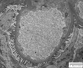
Just think of the life without space and you would understand its importance. Our body is like a tube open from both ends. You may appreciate space in your mouth (oral cavity), throat, nostrils, ears and lungs. In addition to these gross pockets of space there are hollow organs like heart (four chambers are there for blood flow regulation), gall bladder, urinary bladder and uterus (in females). Other examples of space within our body are cranial cavity, visceral cavity and cavities around all vital organs. There are micro-spaces in glandular tissues, alveoli of lungs, blood vessels and nephrons (glomeruli have capillary lumen and urinary space) in kidneys. In some of the renal disorders there are ultrastructural alterations in the areas/volumes of these micro-spaces within the kidneys leading to altered renal physiology and renal function. The figure-1 below illustrates normal urinary space (US) and capillary lumen (CL) or capillary space in a normal kidney; and figure-2 illustrates congestion of capillary lumen (CL) due to deposition of subendothelial deposits (SeD) in a kidney affected by lupus nephritis.

Figure-1: Ultramicrograph of a capillary loop from a normal human kidney illustrating normal urinary space (US) and capillary lumen (CL) with normal thickening of glomerular basement membrane (GBM); Uranyl acetate and Lead citrate stain.

Figure-2: Ultramicrograph of a capillary loop from human kidney affected by lupus nephritis, illustrating congestion of capillary lumen (CL) due to deposition of subendothelial deposits (SeD) with normal urinary space (US) but irregular thickening of glomerular basement membrane GBM); Uranyl acetate and Lead citrate stain.

In the illustration cited above you have seen the alteration in the space within the renal glomerulus. Abdominal tumors, brain tumors, polyps in the uterus, enlargement of spleen and liver, all these lead to functional as well as physiological changes in the body of a patient due to impact on space within the body.