Showing posts with label kidney. Show all posts

Wednesday, February 24, 2010

Diagnosis and Type of Kidney Disease – Investigations and interpretations

Correlation of clinical and laboratory features is must for an accurate diagnosis and type of a kidney disease (renal disease) or glomerulonephritis. An experienced nephrologist can make a diagnosis of glomerulonephritis from thorough history, physical examination, urine examination and microscopy of urinary sediment. The assessment of presenting features of the patient, such as nephritic or nephrotic syndrome is important. However, the decision on the type of glomerulonephritis can not be based on the clinical and laboratory features; as the nephrotic syndrome may occur with any histological glomerulonephritis, and nephritic syndrome is the outcome of proliferative glomerulonephritis. So the ultimate diagnostic tool is renal biopsy and its light and fluorescent microscopy as well as ultrastructural study by electron microscope.

The interpretation of clinical features in the light of histological diagnosis of renal biopsy helps the clinician to detect any systemic disease associated with the renal disease (kidney disease). Majority of the patients with suspected glomerulonephritis need renal biopsy evaluation. However, in children with nephrotic syndrome; if there is no microscopic hematuria (blood in urine) and red cells' or granular casts, renal biopsy procedure may be avoided initially. In patients, who do not respond to steroid therapy; renal biopsy investigation is must. There are around one million glomeruli (1x10⁶ glomeruli) in each kidney and at least 5 glomeruli should be included in the renal biopsy evaluated histologically to achieve a diagnosis of glomerulonephritis.

Radiological and laboratory investigations in glomerulonephritis:

The clinical presentation, urine-analysis and microscopy findings, and presence of a normal upper & lower urinary tract on intravenous pyelography (IVP: a radiological investigation) or ultrasonography without any renal scarring could be indicative of glomerulonephritis, but there could be a need for renal biopsy.

Immune system associated investigations:

Our body is equipped with a multitasking immune system composed on lymphocytes, antibodies and complement system. The immune system always defends our body internally against a variety of infections and pathological conditions; and assessment of its components and abnormal products produced by it helps in diagnostic conclusions. Complement system of our body is composed of 9-components and boosts the body defense in association with cellular components. The blood level of complement components C3, C4 and C1q may be reduced or normal in some renal diseases. Low total serum complement, C3, C4 and C1q levels are observed in glomerulonephritis associated with circulatory immune complex disorders like systemic-lupus erythematosis (SLE), bacterial endocarditis and serum sickness. Normal levels of C4 and C1q but decreased level of C3 is generally observed in membranoproliferative glomerulonephritis (MPGN) and dense deposit disease of the kidney.

Following investigations are considered important to ascertain the diagnosis and type of glomerulonephritis:

Investigations for likely diagnosis of glomerulonephritis:

  • Clinical presentation
  • Urine analysis (proteinuria, hematuria and electrophoresis)
  • Microscopy of urinary sediment
  • Intravenous pyelography (IVP: Radiological investigation)
  • Abdominal ultrasonography.

Investigations for likely type of glomerulonephritis:

  • Estimation of serum complement components' level
  • Detection of circulating immune complexes
  • Detection of auto-antibodies such as anti-nuclear antibodies (ANA), anti-DNA antibodies and anti-glomerular basement membrane antibodies (anti-GBM antibodies)
  • Renal biopsy

Investigations for assessing the implications of glomerulonephritis and monitoring the effect of therapy:

  • Determination of 24 hour urinary protein
  • Determination of level of serum proteins
  • Determination of serum cholesterol and/or lipid profile
  • Determination of serum creatinine, blood urea and serum electrolytes.

Monday, September 28, 2009

IgA Nephropathy as a cause of End Stage Renal Disease

There are a variety of causes of end stage renal disease (ESRD) in teenagers and adults. Immunoglobulin-A (IgA) nephropathy could be a cause of end stage renal disease (ESRD) in around 25% of cases. There are five types of immunoglobulins in our body for protection against microorganisms and IgA provides defence at mucous membranes. Colostrum and breast milk are rich sources of IgA and protect us during infancy through breast-feeding. However, later in life, chronic mucosal inflammation (inflammation of respiratory, oral, or gastrointestinal mucous membranes) may lead to IgA-nephropathy (IgAN). Viral (including HIV), bacterial, yeast and parasitic infections have been found to be associated with IgAN. Environmental and food antigens have also been implicated in IgAN as these may mimic molecular structure of microbial antigens and lead to excessive IgA production, aggregation and breakdown of mucosal barrier. Patients affected by IgAN may present with hematuria (blood in urine) and/or proteinuria (protein in urine) with or without rise in serum creatinine. The most common initial symptom in children is microscopic hematuria. Some adults may present with acute or chronic renal failure.

IgA nephropathy is a common nephropathy, which could be detected on renal (kidney) biopsy evaluation through light and fluorescence microscopy. However, electron microscopic study of renal biopsy acts as a diagnostic adjunct as the location of immune complexes in the renal glomerulus could be pronounced on electron micrographs. Figures 1 and 2 are the electron micrographs from a proven case of IgAN, illustrating mesangial deposits of IgA.

Figure-1: Electron micrograph of an area of glomerulus of a case of IgAN showing electron dense deposits (D) in the mesangial (Mes) area. Glomerular basement membrane (GBM), capillary lumen (CL), podocyte or epithelial cell (EpC) and urinary space (US) are also exhibited; Original Magnification 4600x.

Figure-2: Electron micrograph of an area of glomerulus of a case of IgAN showing electron dense deposits (D) in the mesangial (Mes) area. Glomerular basement membrane (GBM), capillary lumen (CL), podocyte or epithelial cell (EpC) and urinary space (US) are also exhibited; Original Magnification 6000x.

The pathology of IgAN may be variable depending on underlying cause. Mesangioproliferative glomerulonephritis is the most common pattern in many renal biopsies; however, glomeruli may appear normal on light microscopy in some of the cases. Renal biopsies in a few cases may also show crescent formation in occasional glomeruli. Diagnosis of IgA nephropathy is established by direct immunofluorescence technique on renal biopsies and the pattern may be dominant or co-dominant for IgA staining. The incidence of ESRD has been found to be high in patients presenting with >1g/day proteinuria with increased level of serum creatinine as compared to those having proteinuria <1g/day with increased level of serum creatinine. Pathogenesis of IgAN is very complex. A variety of underlying diseases including hepato-biliary disease can be associated with IgA nephropathy. Defective detection and clearance by liver of polymeric immune complexes of IgA (IgA1) due to abnormal galactosylation of O-linked glycans is probably the major cause of IgAN in addition to loss of mucosal barrier and chronic mucosal inflammation. Recurrent tonsillitis may also lead to IgA nephropathy and tonsillectomy may be helpful in these cases to remove the mucosal foci of infection. Optimal treatment of tonsillitis and other oromucosal infections with antibiotics along with conventional treatment of IgAN would be helpful to put brakes on the progression of IgA nephropathy. Patients with acute or chronic renal failure due to advanced stage of IgAN may need hemodialysis or renal transplantation. Use of anti-oxidants and fish oil as food supplements in some cases of IgA nephropathy have been found beneficial.

Monday, August 3, 2009

Renal Transplantation and Immune Profiling

Organ transplantation is analogous to blood transfusion and we need to detect and match the tissue antigens of the donor and the recipient before transplantation of an organ, say kidney. Tissue antigens are known as human leucocyte antigens (HLA). There are four loci called A, B, C and D on the 6th chromosome, which govern these tissue antigens or HLA. We inherit one gene (each gene has sub-genes) each on each locus from our mother and father. There is antigenic polymorphism at each locus (A, B, C, and D). Unless the kidney donor and the recipient (patient) are identical twins, a 100% match of these HLA is not possible. There is 50% match of HLA amongst parents and children, and the siblings. Unrelated donor and recipient may also have 50% matching of tissue antigens or HLA. The participation of immune mechanisms in allogenic kidney transplant begins with the identification and appropriate reaction to the donor organ, by the recipient, depending on the degree of HLA mismatch. Immunosuppressive therapeutic protocols are prescribed for the adoption and survival of grafted/transplanted kidney. There is very complex immune pathway in our body involving antigen presenting cells and T & B cells (Lymphocytes), which get activated and lead to injury of the target cells. The intragraft cell trafficking and their effector mechanisms may have serious implications. Post transplant immune profiling is a way of monitoring the allograft function and to elucidate pathogenic mechanisms and molecular pathways causing tissue injury and disease.

Transplant tolerance could only be achieved through sincere compliance of immunosuppressive therapy. The immune system of the recipient following renal transplantation, though challenged by the exposure to donor antigens to initiate an early sub-clinical or acute rejection process, attempts to regulate the inflammatory processes or maintain homoeostasis in the body. The acute rejection may be cell or antibody mediated. The transplant tolerance is defined as maintenance of stable allograft function without clinical evidence of immunosuppression. There are many therapeutic approaches to achieve the transplant tolerance, however, the best one is donor specific transfusion or hematopoietic cell infusion. Almost all the transplant recipients have to depend on a variety of immunosuppressive protocols to ward of any chance of allograft rejection.

Thursday, July 30, 2009

End Stage Renal Disease and Renal Transplantation

Chronic glomerulonephritis, diabetic nephropathy, chronic tubulointerstitial disease, benign nephrosclerosis and polycystic kidney disease are the major causes of end stage renal disease (ESRD) and renal failure. Patients with ESRD exhibit a variety of abnormalities in their autonomic functions. Precise mechanisms of evaluating autonomic functions have revealed abnormalities in efferent parasympathetic pathway and baroreceptor sensitivity in patients with end stage renal disease. An increase in expiration-inspiration, lying standing and valsalva ratios, and baroreceptor sensitivity slope have been well documented in ESRD. Uremic patients with ESRD respond poorly to antihypertensive drugs as compared to otherwise healthy controls. Renal involvement in multiple myeloma is an other cause of ESRD and renal failure. Dialysis is an adoptive procedure in patients having end stage renal disease and ultimate surgical measure is renal (kidney) transplantation. Adequate dialysis in patients with ESRD reverses the elevated levels of urea, creatinine and electrolytes in blood.

Though renal transplantation is must in patients with ESRD, but it needs a lot of medication and post transplantation care for the successful adoption and survival of renal allograft. Systemic fungal infections (cryptococcosis, mucuromycosis, candidiasis, aspergillosis and mixed infections) have been documented after renal transplantation. Though these infections are treatable but may complicate the post operative care as additional medication will be required in addition to immunosuppressive therapy. High incidence of tuberculosis has also been observed in recipients of renal transplant along with viral infections like BK virus and cytomegalovirus (CMV). Adverse impact of pre-transplant polyoma virus (BK virus) infection on the graft survival has also been documented. Molecular technology has been developed for the early detection and identification of these viruses from the time of renal transplantation onwards by using protocol biopsies from the grafted kidney.

Tuesday, June 30, 2009

The Space Within and Outside Our Body

The space has a great role in our life. Our body is composed of five basic components: the earth, water, air, fire (heat or temperature) and the sky or space. The space within and outside our body is must for the existence of life. The outer space is composed of air (mixture of gases), vapours, finer particles, microorganisms, radiation, light, cold and heat. The composition of environment influences our breathing, metabolism and physiology. Our body reacts in a variety of ways to the external space and the environment possessed by it. In fact the particles floating in the air or transmitted through it may cause allergic reactions, infections, hot or cold skin burns or even skin cancer. All activities of human beings or animals are space oriented.

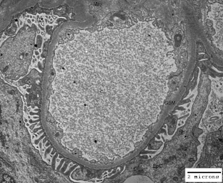
Just think of the life without space and you would understand its importance. Our body is like a tube open from both ends. You may appreciate space in your mouth (oral cavity), throat, nostrils, ears and lungs. In addition to these gross pockets of space there are hollow organs like heart (four chambers are there for blood flow regulation), gall bladder, urinary bladder and uterus (in females). Other examples of space within our body are cranial cavity, visceral cavity and cavities around all vital organs. There are micro-spaces in glandular tissues, alveoli of lungs, blood vessels and nephrons (glomeruli have capillary lumen and urinary space) in kidneys. In some of the renal disorders there are ultrastructural alterations in the areas/volumes of these micro-spaces within the kidneys leading to altered renal physiology and renal function. The figure-1 below illustrates normal urinary space (US) and capillary lumen (CL) or capillary space in a normal kidney; and figure-2 illustrates congestion of capillary lumen (CL) due to deposition of subendothelial deposits (SeD) in a kidney affected by lupus nephritis.

Figure-1: Ultramicrograph of a capillary loop from a normal human kidney illustrating normal urinary space (US) and capillary lumen (CL) with normal thickening of glomerular basement membrane (GBM); Uranyl acetate and Lead citrate stain.

Figure-2: Ultramicrograph of a capillary loop from human kidney affected by lupus nephritis, illustrating congestion of capillary lumen (CL) due to deposition of subendothelial deposits (SeD) with normal urinary space (US) but irregular thickening of glomerular basement membrane GBM); Uranyl acetate and Lead citrate stain.

In the illustration cited above you have seen the alteration in the space within the renal glomerulus. Abdominal tumors, brain tumors, polyps in the uterus, enlargement of spleen and liver, all these lead to functional as well as physiological changes in the body of a patient due to impact on space within the body.

Tuesday, December 23, 2008

What Could Be The Cause Of Swelling On Face

The swelling on face or facial edema should be taken seriously if there is no history of insect bite, wasp sting or honey bee sting and when it is after a throat infection. The swelling on face or facial edema could be due to renal disorder (kidney disease). If on routine examination of urine of the patient, excretion of albumin or protein is detected; there is a need to consult a nephrologist for proper investigations. Blood biochemistry for blood urea, serum creatinine, serum proteins, serum electrophoresis, urine electrophoresis and 24-hour urinary protein should be done. Excretion of protein in 24-hours through urine will help the physician to assess the loss of proteins and possible course of action. Urine electrophoresis would show the type of protein being excreted in the urine. In a patient with nephrotic syndrome, serum electrophoresis would show hypoalbuminemia (low level of albumin in blood), hypogammaglobulinemia (low level of globulins in blood) and raised alpha-2 (a-2) globulin, and urine electrophoresis may show albuminuria (excretion of albumin in urine) or non-selective proteinuria (excretion of almost all the fractions of serum proteins in urine). Total serum protein and its fractions like albumin and globulin would show the altered albumin-globulin ratio. The normal albumin-globulin ratio (Albumin/Globulin) is 3:1 and it may be reversed in patients with swelling on face due to kidney disease.

The swelling on face or facial edema is directly associated with albuminuria (excretion of albumin in urine) and salt retention. The loss of blood albumin through urine hinders the return of fluid from the tissues into the blood and may thus lead to development of edema. It is well known that 68 to 70% weight of our body is due to water content in the blood and tissues. Around 12 to 14% of the total water volume of our body is in the blood and the rest is present in the tissues of the body. There is direct correlation between albuminuria (excretion of albumin in urine) and edema. Retention of Chloride is also a common accompaniment of edema. However, there may not be any retention of Chloride in majority of the cases with edema. The edema is perhaps the greatest problem confronting the students of nephrology. Pathological lesions in the kidney need to be evaluated microscopically through renal biopsy examination. Blood urea and serum creatinine may be normal. There may be salt retention without edema and edema without salt retention. The Chloride may collect in watery subcutaneous tissue due to some external factors also without involvement of any renal lesion.

Two forms of swelling on face or facial edema could be recognized and these are called nephritic edema and nephrotic edema. In nephritic edema the protein content of the edema fluid is over 1 gram/dl whereas in nephrotic edema the protein content of the edema fluid is always less than 0.1 gram/dl. Nephritic edema occurs in acute glomerulonephritis. The capillaries in the subcutaneous tissue become more permeable leading to leakage of proteins in the extra cellular fluid. Nephrotic edema occurs in the wet nephritis or second stage of nephritis, in nephrosis and also in renal amyloidosis. The edema is caused due to the great fall in the osmotic pressure of the blood due to constant loss of protein in urine; so, the fluid from the blood vessels escapes into the tissues in an effort to correct the viscosity of blood plasma.

Friday, November 21, 2008

Essential Ions Of Our Body For Sustaining Life

Our life is sustained by complex interaction of inorganic and organic substances; and the water serves as the supportive medium and vehicle for the elements of life. There are nine types of most essential ions of our body which play a dynamic role in supporting and sustaining health and life. Out of nine, five are positively charged ions and four are negatively charged ions. The positively charged ions are called cations as these collect at the negative electrode or cathode during electrolysis; these are Na+ (Sodium ion), K+ (Potassium ion), Ca++ (Calcium ion), Mg++ (Magnesium ion) and H+ (Hydrogen ion). The negatively charged ions are called anions as these collect at positive electrode or anode during electrolysis; these are Cl- (Chloride ion), HCO3- (Bicarbonate ion), PO43- (Phosphate ion) and OH- (Hydroxyl ion). The genius, Sir Humphrey Davy discovered in the second decade of the 19th Century that the passage of current through the aqueous solution of inorganic compounds dissociated them into positively charged and negatively charged parts. That was the beginning of a field which today has the great utility in the life sciences and industry. The Sodium (Na+), Chloride (Cl-), Potassium (K+) and Bicarbonate (HCO3-) are called principle electrolytes and are present in the blood and various body fluids. Potassium (K+) is essential for heart function. The concentration of electrolytes is expressed in milli-Equivalents (mEq) per litre. In the plasma of our blood the normal concentration of Sodium is around 145 mEq/litre; Potassium ranges from 3 to 5 mEq/litre; Chloride is around 100 mEq/litre and Bicarbonate is around 30 mEq/litre. Hydrogen ions (H+) hydroxyl ions (OH-), Bicarbonate ions (HCO3-) and Phosphate ions (PO43-) govern the acid-base balance. Our kidneys play a vital role in maintaining the electrolyte balance and acid-base balance. The total sum of electrolytes also determines the osmotic pressure of intracellular and extracellular or interstitial body fluids. The osmotic pressure is a physical force and expressed in osmols (Osm) or milliosmols (mOsm). One mEq of monovalent ions would exert one mOsm and one mEq of divalent ions would exert two mOsm of osmotic pressure. Osmotic pressure regulates the movement of water from a compartment of lower concentration of electrolytes or ions to a compartment of higher concentration of electrolytes or ions.

Daily requirement of salts and minerals of and adult person is 3 to 5 grams. Common salt (Sodium Chloride) is a source of Sodium and Chloride ions. Rock salt also provides Potassium ions as it contains some Potassium Chloride in addition Sodium Chloride. Fruits, vegetables and animal products are rich in salts and minerals. Excess of salts taken as food additives are excreted in urine by kidneys. One may loose electrolytes and water due to excessive sweating, continued vomiting or profuse diarrhea resulting in dehydration. To offset the ill effects of dehydration and to correct the lost electrolytes we should drink the solution of oral rehydration salts that contains Sodium Chloride, Potassium Chloride, Sodium Citrate and Glucose in optimal proportions.

Tuesday, July 8, 2008

Internal Environment & Renal Physiology

Internal Environment: The body is made up of organs and tissues each composed of cells and fibers that constitute their histology. Water with its solvents needed for the health of the cells is termed as body fluid and this fluid is partly inside and partly outside the cells. Water constitutes about 70 per cent of fat free body weight. Water is the fabric of everything that lives. The body of a baby contains mostly water, whilst the old man or woman shrivels up like a wilted plant. We are completely immersed in water during the first nine months of life in the mother's womb. Water is involved in the health, disease and death. Loss of water leads to dehydration and may cause death if not corrected. Retention of water leads to edema and may cause death if remedial action is not taken. For each cell in the body the same conditions prevail as for the single-celled creatures fixed on the bed of a flowing stream which brings their food and oxygen and carries away their waste material. In our body water or the body fluid is controlled in the two major compartments: (1) intracellular compartment for intracellular fluid (2) extracellular compartment for extracellular fluid. The extracellular fluid is of two subtype (1) interstitial fluid (2) blood plasma. Intracellular fluid makes up about 40 to 50 per cent of the body weight and bulk of it being contained in muscles. Extracellular fluid represents about 20 per cent of the body weight, of which 15 per cent is interstitial fluid including lymph and 5 per cent constitute the blood plasma. The interstitial fluid constitutes the real internal environment. It is the adjustable segment in the total water content of the body. Its volume and solutes are regulated by the kidneys, lungs, endocrine glands, and are influenced by sweat glands and gastrointestinal tract. The blood plasma is in equilibrium with the interstitial fluid. Both the vascular and intracellular compartments contain a lot of protein. The normal intake of water in an adult is about 2500 ml. About 2100-2200 ml of this is taken by mouth as food and pure water and rest is the endogenous water from cellular oxidation. Renal Physiology: The word renal pertains to kidney in medical terminology. Water regulation in our body is achieved by water loss through four routes: (1) intestine (2) lungs (3) skin and (4) kidneys. Kidneys play a major role in water regulation as these excrete 50 to 70 per cent of excess water. Major functions of kidneys are: (a) excretion/elimination of excess water from body (b) excretion/elimination of waste products of metabolism e.g. urea and creatinine (c) excretion/elimination of foreign substances such as drugs (d) retention of substances necessary for normal body functions (major substances are proteins, amino acids and glucose) (e) regulation of electrolyte balance and osmotic pressure of the body fluids. Sodium ions, potassium ions, bicarbonate ions and chloride ions are major electrolytes. Urea is the main product of protein metabolism in the body. Removal of amino groups from amino-acids, from which urea is formed, takes place in the liver. Urine urea estimations are most commonly carried out as part of renal efficiency tests. A high concentration of urea in the urine shows that the kidneys possess a good concentrating power. However, in cases where there is increased blood urea due to non-renal or pre-renal factors, urine urea may be quite high. On an average concentration of urea in urine should be 2.0 per cent over the day. The total urea excretion in an adult is about 30 grams daily. At least 1500 ml of water must be excreted by kidneys daily to carry the solids which have to be eliminated. There is a pair of kidneys in our body to accomplish the above task. The kidney is the organ concerned with the regulation of the volume and composition of body fluids. Each kidney contains over 1000,000 functional units called nephrons. Each nephron consists of (a) glomerulus with its afferent and efferent arteriole (b) proximal convoluted tubule (c) loop of Henle (d) distal convoluted tubule and (e) collecting tubule. The structure of the glomerulus is that of a filtration mechanism. The afferent arteriole divides into 3 or 4 branches, which gives the lobulated appearance to the glomerular tuft. Each branch gives rise to 40 to 50 capillary loops, which probably do not anastomose with one another. The diameter of efferent arteriole is only half of that of afferent arteriole and the efferent arteriole splits up into a huge network of capillaries containing blood that is highly viscous by reason of the preceding loss of water. The viscous blood moves slowly, so raises the pressure in the glomerular tuft and thus facilitate filtration. The glomerular tuft consists of four main components: (i) the endothelium lining the capillaries (ii) the basement membrane which separates the endothelium from (iii) the epithelium and (iv) the mesangium. Mechanism of Renal Function: Every minute about 1000 ml of blood containing about 500 ml of plasma flows through the glomeruli of kidneys and about 100 ml of it is filtered out as raw-urine called glomerular filtrate. The plasma containing all the salts, glucose and other small substances is filtered in the glomerular filtrate. The cells and plasma proteins are too big to pass through the pores of the filter and stay behind in the blood stream. The glomerular filtrate then passes through the real tubules and 85% of it is absorbed automatically by the proximal tubules, where essential substances are reabsorbed. The fate of remaining 15% depends upon the degree of further reabsorption of water in the distal tubules. The reabsorption is controlled by antidiuretic hormone (ADH) released from the posterior pituitary gland (an endocrine gland). Loss of ADH results in defective reabsorption of water in the distal tubules and causes diabetes insipidus. An increase in the electrolyte osmotic pressure (osmolality) of the extracellular fluids results in an increased release of ADH with an increased water reabsorption in distal tubules. Conversely any decrease in the osmolality will lead to opposite effect. This complex controlling mechanism is termed as neurohypophysial-renal axis. Just as the electrolytic osmotic pressure of the extracellular fluid is controlled by the ADH, the volume of that fluid is controlled by aldosterone from the adrenal gland. The ADH regulates the retention or excretion of water and the aldosterone regulates the reabsorption of sodium and thus the retention of water. Thus the secretion of urine is accomplished in three steps: (a) glomerular filtration. (b) tubular reabsorption and (c) tubular secretion. By comparing the amount filtered by the glomeruli per day with the amount usually excreted in the urine we can see how selective is renal function? Daily about 150 liters of water is filtered and about 1500 ml is excreted; about 750 grams of salts are filtered and 15 grams are excreted; about 150 grams of glucose are filtered and no amount is excreted and about 50 grams of urea are filtered and about 30 grams excreted.