Showing posts with label GBM. Show all posts

Tuesday, June 30, 2009

The Space Within and Outside Our Body

The space has a great role in our life. Our body is composed of five basic components: the earth, water, air, fire (heat or temperature) and the sky or space. The space within and outside our body is must for the existence of life. The outer space is composed of air (mixture of gases), vapours, finer particles, microorganisms, radiation, light, cold and heat. The composition of environment influences our breathing, metabolism and physiology. Our body reacts in a variety of ways to the external space and the environment possessed by it. In fact the particles floating in the air or transmitted through it may cause allergic reactions, infections, hot or cold skin burns or even skin cancer. All activities of human beings or animals are space oriented.

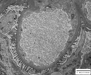
Just think of the life without space and you would understand its importance. Our body is like a tube open from both ends. You may appreciate space in your mouth (oral cavity), throat, nostrils, ears and lungs. In addition to these gross pockets of space there are hollow organs like heart (four chambers are there for blood flow regulation), gall bladder, urinary bladder and uterus (in females). Other examples of space within our body are cranial cavity, visceral cavity and cavities around all vital organs. There are micro-spaces in glandular tissues, alveoli of lungs, blood vessels and nephrons (glomeruli have capillary lumen and urinary space) in kidneys. In some of the renal disorders there are ultrastructural alterations in the areas/volumes of these micro-spaces within the kidneys leading to altered renal physiology and renal function. The figure-1 below illustrates normal urinary space (US) and capillary lumen (CL) or capillary space in a normal kidney; and figure-2 illustrates congestion of capillary lumen (CL) due to deposition of subendothelial deposits (SeD) in a kidney affected by lupus nephritis.

Figure-1: Ultramicrograph of a capillary loop from a normal human kidney illustrating normal urinary space (US) and capillary lumen (CL) with normal thickening of glomerular basement membrane (GBM); Uranyl acetate and Lead citrate stain.

Figure-2: Ultramicrograph of a capillary loop from human kidney affected by lupus nephritis, illustrating congestion of capillary lumen (CL) due to deposition of subendothelial deposits (SeD) with normal urinary space (US) but irregular thickening of glomerular basement membrane GBM); Uranyl acetate and Lead citrate stain.

In the illustration cited above you have seen the alteration in the space within the renal glomerulus. Abdominal tumors, brain tumors, polyps in the uterus, enlargement of spleen and liver, all these lead to functional as well as physiological changes in the body of a patient due to impact on space within the body.
Thursday, January 22, 2009

Acute Glomerulonephritis

The term acute glomerulonephritis is used by clinicians as well as pathologists to describe the sudden onset of kidney disease. Irrespective of the cause, there would be enlargement of a kidney or kidneys and the capsule around the kidney is strained and stretched. Ultrasonography is always helpful to ascertain the size of kidneys. Histological examination of kidney biopsy would present densely cellular glomerular tuft with polymorphonuclear cells (a type of white blood cells) in the glomerular capillaries. Accumulation of leukocytes (white blood cells) in the glomerular capillaries with swelling and proliferation of the vascular endothelium (inner lining of capillaries) is rapidly followed by edema (swelling) and mesangial proliferation (enlargement of inner stalk of a cluster of glomerular capillaries)leading to capillary ischemia (poor blood supply).

The glomerular basement membrane (GBM) is the other important component affected by acute glomerulonephritis. By electron microscopic study of kidney biopsy, electron dense deposits of antigen-antibody complexes could be revealed in and around the glomerular basement membrane. The ultrastructural features of normal GBM have been depicted in the figure-1 and the figure-2 is from a case affected by acute glomerulonephritis.

Figure-1: Electron micrograph of a capillary loop of glomerular tuft showing normal features; CL: capillary lumen, US; urinary space, En: endothelium, GBM: glomerular basement membrane, EpC: epithelial cell or podocyte.

Figure-2: Electron micrograph of a capillary loop of glomerular tuft from a case affected by acute glomerulonephritis is showing sub-epithelial deposits; CL: capillary lumen, US; urinary space, GBM: glomerular basement membrane, dep: deposits on sub-epithelial site of GBM.

The tubules in the kidney may show slight degenerative changes. The degree of degenerative changes in the tubules depends on the extent of the glomerular obstruction. The interstitial tissue and the blood vessels are observed to be normal on the histological study of kidney biopsy.

Thursday, January 8, 2009

Azotemic or Hydropic Glomerulonephritis

The high concentration of non protein nitrogen (NPN) in the blood of a patient, mainly due to elevated level of urea is termed as azotemia. Elevated level of urea in blood is termed as uremia and it may be due to renal abnormality or due to other health problems like dehydration and excessive burns on the body. The glomerulonephritis (inflammation of glomeruli of kidneys) may be azotemic or hydropic type depending on the nature of glomerular lesions. There may be marked narrowing of the glomerular capillaries leading to azotemia with renal insufficiency and hypertension. On the other hand the status of glomerular capillaries may remain normal but there could be an increase in the permeability of glomerular basement membrane (GBM), the filtration barrier of kidneys. The clinical picture would be hydropic (accumulation of water in the tissues of the body) in character. The hydropic glomerulonephritis is clinically represented with edema associated with hypoproteinemia (low level of protein in blood) and hyperlipemia (high concentration of lipids or cholesterol in blood).

A child or an adult affected with fever or some other acute disease finds the increase in the daily output of urine. There may be tinge of blood in the urine of the patient affected by glomerulonephritis. Sudden appearance of features like puffiness or swelling on face (facial edema), ankles and hands after any acute disease needs expert medical attention and medication. The urine of such patients would show notable excretion of albumin. The patient may be less perspiring with dry skin. The pulse could be full and hard. The loin pain and a feeling of heaviness in the lower abdominal region are the associated symptoms. The polyuria (excessive output of urine) is probably compensatory action of affected kidneys to flush out solid wastes of metabolism form the body. An effective therapy would definitely reverse the associated symptoms and lesions.